WB result of CD19 Mouse mAb
Primary antibody: CD19 Mouse mAb at 1/1000 dilution
Lane 1: untreated Raji whole cell lysate 20 µg
Lane 2: Raji treated with PNGase F whole cell lysate 20 µg
Secondary antibody: Goat Anti-mouse IgG, (H+L), HRP conjugated at 1/10000 dilution
Predicted MW: 61 kDa
Observed MW: 95 kDa
CD19 Mouse mAb (S-1039-46)
CD19 Mouse mAb (S-1039-46)
Price:
Regular price
$100 USD
Regular price
Sale price
$100 USD
Unit price
per
For shipping services or bulk orders, you may request a quotation.
Secure checkout with

View full details
Product Details
Product Details
Product Specification
| Host | Mouse |
| Antigen | CD19 |
| Synonyms | B-lymphocyte antigen CD19, LE-CD19, B-lymphocyte surface antigen B4, Differentiation antigen CD19, T-cell surface antigen Leu-12 |
| Immunogen | Recombinant Protein |
| Location | Cell membrane |
| Accession | P15391 |
| Clone Number | S-1039-46 |
| Antibody Type | Mouse mAb |
| Isotype | IgG1,k |
| Application | WB, IHC-P, ICC, FCM, IF |
| Reactivity | Hu |
| Purification | Protein G |
| Concentration | 2 mg/ml |
| Conjugation | Unconjugated |
| Physical Appearance | Liquid |
| Storage Buffer | PBS, 40% Glycerol, 0.05% BSA, 0.03% Proclin 300 |
| Stability & Storage | 12 months from date of receipt / reconstitution, -20 °C as supplied |
Dilution
| application | dilution | species |
| WB | 1:1000 | |
| IHC-P | 1:500-1:1000 | |
| ICC | 1:500 | |
| FCM | 1:2000 | |
| IF | 1:500 |
Background
The CD19 antigen plays an important role in clinical oncology. In normal cells, it is the most ubiquitously expressed protein in the B lymphocyte lineage. CD19 expression is induced at the point of B lineage commitment during the differentiation of the hematopoietic stem cell, and its expression continues through preB and mature B cell differentiation until it is finally down-regulated during terminal differentiation into plasma cells. CD19 expression is maintained in B-lineage cells that have undergone neoplastic transformation, and therefore CD19 is useful in diagnosis of leukemias and lymphomas using monoclonal antibodies (mAbs) and flow cytometry [PMID: 8528044].
Picture
Picture
Western Blot
FC
Flow cytometric analysis of human PBMC (human peripheral blood mononuclear cell) labelling CD19 antibody at 1/2000 (0.1 μg) dilution (Right) compared with a Mouse monoclonal IgG isotype control (Left). Goat Anti - Mouse IgG Alexa Fluor® 488 was used as the secondary antibody. Then cells were stained with CD3 - Alexa Fluor® 647 separately. Gated on total viable cells.
Immunohistochemistry
IHC shows positive staining in paraffin-embedded human spleen. Anti-CD19 antibody was used at 1/1000 dilution, followed by a HRP Polymer for Mouse & Rabbit IgG (ready to use). Counterstained with hematoxylin. Heat mediated antigen retrieval with Tris/EDTA buffer pH9.0 was performed before commencing with IHC staining protocol.
IHC shows positive staining in paraffin-embedded human tonsil. Anti-CD19 antibody was used at 1/500 dilution, followed by a HRP Polymer for Mouse & Rabbit IgG (ready to use). Counterstained with hematoxylin. Heat mediated antigen retrieval with Tris/EDTA buffer pH9.0 was performed before commencing with IHC staining protocol.
IHC shows positive staining in paraffin-embedded human diffuse large B-cell lymphoma. Anti-CD19 antibody was used at 1/500 dilution, followed by a HRP Polymer for Mouse & Rabbit IgG (ready to use). Counterstained with hematoxylin. Heat mediated antigen retrieval with Tris/EDTA buffer pH9.0 was performed before commencing with IHC staining protocol.
Negative control: IHC shows negative staining in paraffin-embedded human anaplastic large cell lymphoma. Anti-CD19 antibody was used at 1/500 dilution, followed by a HRP Polymer for Mouse & Rabbit IgG (ready to use). Counterstained with hematoxylin. Heat mediated antigen retrieval with Tris/EDTA buffer pH9.0 was performed before commencing with IHC staining protocol.
Immunocytochemistry
ICC shows positive staining in Raji cells (top panel) and negative staining in Jurkat cells (below panel). Anti-CD19 antibody was used at 1/500 dilution (Green) and incubated overnight at 4°C. Goat polyclonal Antibody to Mouse IgG - H&L (Alexa Fluor® 488) was used as secondary antibody at 1/1000 dilution. The cells were fixed with 100% ice-cold methanol and permeabilized with 0.1% PBS-Triton X-100. Nuclei were counterstained with DAPI (Blue). Counterstain with tubulin (Red).
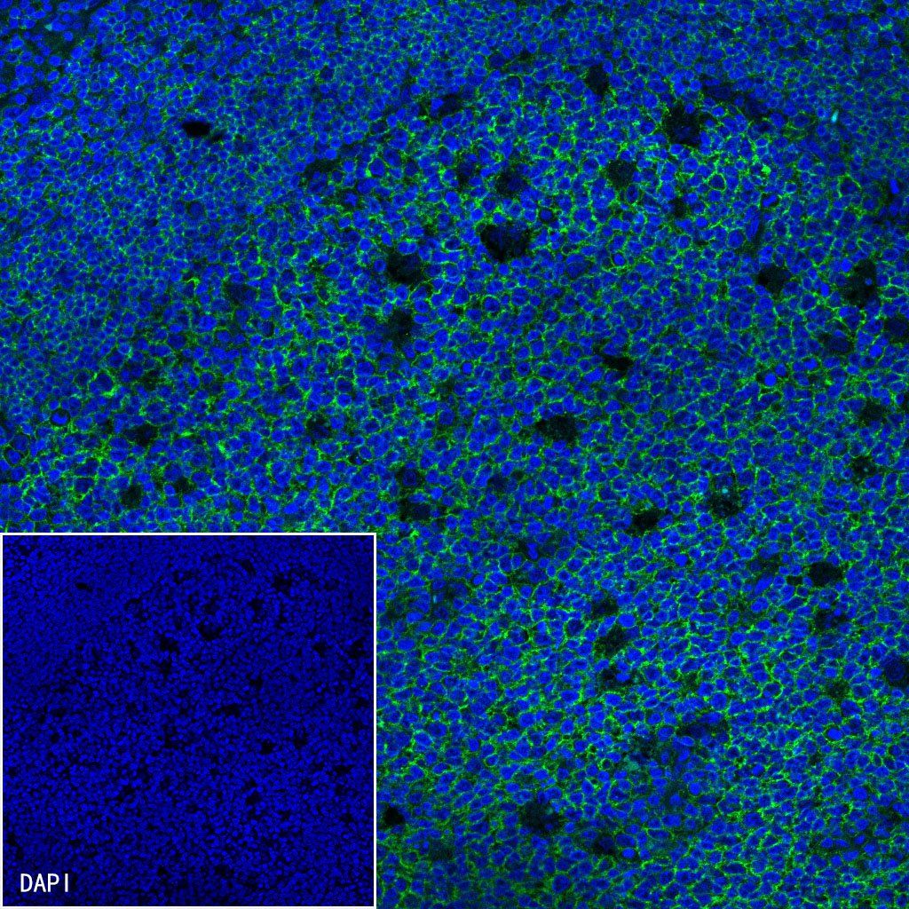
Immunofluorescence
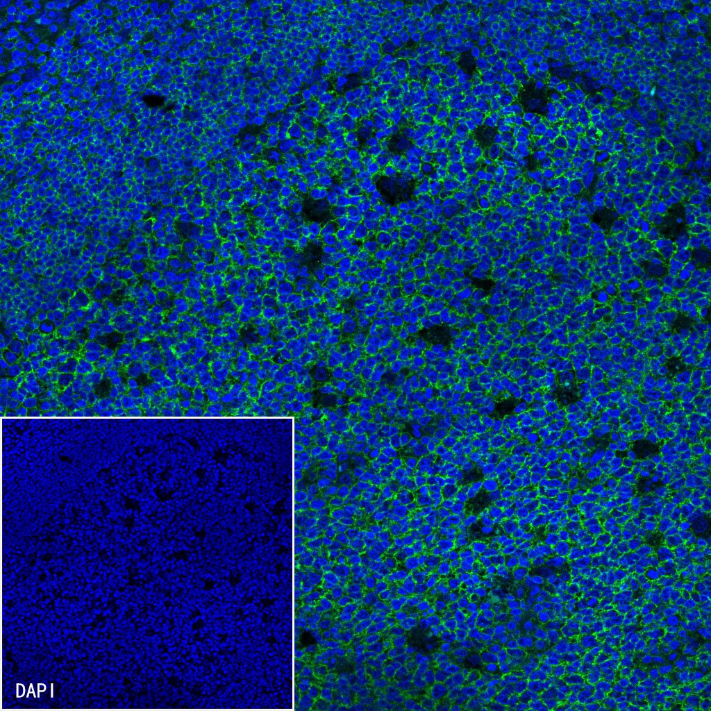
IF shows positive staining in paraffin-embedded human tonsil. Anti-CD19 antibody was used at 1/500 dilution (Green) and incubated overnight at 4°C. Goat polyclonal Antibody to Mouse IgG - H&L (Alexa Fluor® 488) was used as secondary antibody at 1/1000 dilution. Counterstained with DAPI (Blue). Heat mediated antigen retrieval with EDTA buffer pH9.0 was performed before commencing with IF staining protocol.