WB result of CD137 Rabbit mAb
Primary antibody: CD137 Rabbit mAb at 1/1000 dilution
Lane 1: untreated mouse splenocytes whole cell lysate 20 µg
Lane 2: mouse splenocytes treated with 2.5 µg/ml Concanavalin A for 72 hr whole cell lysate 20 µg
Lane 3: CTLL-2 whole cell lysate 20 µg
Secondary antibody: Goat Anti-Rabbit IgG, (H+L), HRP conjugated at 1/10000 dilution
Predicted MW: 28 kDa
Observed MW: 21~25 kDa, 35~40 kDa
(This blot was developed with high sensitivity substrate)
CD137 Recombinant Rabbit mAb (S-547-3)
CD137 Recombinant Rabbit mAb (S-547-3)
Price:
Regular price
$100 USD
Regular price
Sale price
$100 USD
Unit price
per
For shipping services or bulk orders, you may request a quotation.
Secure checkout with

View full details
Product Details
Product Details
Product Specification
| Host | Rabbit |
| Antigen | CD137 |
| Synonyms | Tumor necrosis factor receptor superfamily member 9, 4-1BB ligand receptor, T-cell antigen 4-1BB, Tnfrsf9, Ila, Ly63 |
| Immunogen | Synthetic Peptide |
| Location | Cell membrane |
| Accession | P20334 |
| Clone Number | S-547-3 |
| Antibody Type | Recombinant mAb |
| Isotype | IgG |
| Application | WB, IHC-P, ICC, FCM, IP |
| Reactivity | Ms |
| Purification | Protein A |
| Concentration | 0.5 mg/ml |
| Conjugation | Unconjugated |
| Physical Appearance | Liquid |
| Storage Buffer | PBS, 40% Glycerol, 0.05% BSA, 0.03% Proclin 300 |
| Stability & Storage | 12 months from date of receipt / reconstitution, -20 °C as supplied |
Dilution
| application | dilution | species |
| WB | 1:1000 | null |
| IP | 1:50 | null |
| IHC-P | 1:250 | null |
| ICC | 1:500 | null |
| FCM | 1:500 | null |
Background
CD137, a member of the tumor necrosis factor (TNF) receptor family, is a type 1 transmembrane protein, expressed on surfaces of leukocytes and non-immune cells. CD137 is only expressed on the cell surface after T cell activation. When T cells are activated by Antigen Presenting Cells (APCs), CD137 becomes embedded in CD4+ and CD8+ T cells. CD137 is a costimulatory molecule functioning to stimulate T cell proliferation, dendritic cell maturation, and promotion of B cell antibody secretion. CD137 is also involved in cancer having been found upregulated in cancerous cell lines. CD137/ligand stimulation has been found to lead to stronger anti-tumor responses due to cytotoxic T cell activation and is being examined as a possible anticancer therapy.
Picture
Picture
Western Blot
FC
Flow cytometric analysis of EL4 (Mouse lymphoma T lymphocyte, left) / CTLL-2 (Mouse T lymphocyte, right) cells labelling CD137 antibody at 1/500 dilution (0.1 μg)/ (Red) compared with a Rabbit monoclonal IgG (Black) isotype control and an unlabelled control (cells without incubation with primary antibody and secondary antibody) (Blue). Goat Anti - Rabbit IgG Alexa Fluor® 488 was used as the secondary antibody.
Negative control: EL4
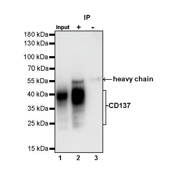
IP
CD137 Rabbit mAb at 1/50 dilution (1 µg) immunoprecipitating CD137 in 0.4 mg mouse splenocytes treated with 2.5 µg/ml Concanavalin A for 72 hr whole cell lysate.
Western blot was performed on the immunoprecipitate using CD137 Rabbit mAb at 1/1000 dilution.
Secondary antibody (HRP) for IP was used at 1/400 dilution.
Lane 1: mouse splenocytes treated with 2.5 µg/ml Concanavalin A for 72 hr whole cell lysate 20 µg (Input)
Lane 2: CD137 Rabbit mAb IP in mouse splenocytes treated with 2.5 µg/ml Concanavalin A for 72 hr whole cell lysate
Lane 3: Rabbit monoclonal IgG IP in mouse splenocytes treated with 2.5 µg/ml Concanavalin A for 72 hr whole cell lysate
Predicted MW: 28 kDa
Observed MW: 21~25 kDa, 35~40 kDa
Immunohistochemistry
IHC shows positive staining in paraffin-embedded mouse spleen. Anti-CD137 antibody was used at 1/250 dilution, followed by a HRP Polymer for Mouse & Rabbit IgG (ready to use). Counterstained with hematoxylin. Heat mediated antigen retrieval with Tris/EDTA buffer pH9.0 was performed before commencing with IHC staining protocol.
IHC shows positive staining in paraffin-embedded mouse thymus. Anti-CD137 antibody was used at 1/250 dilution, followed by a HRP Polymer for Mouse & Rabbit IgG (ready to use). Counterstained with hematoxylin. Heat mediated antigen retrieval with Tris/EDTA buffer pH9.0 was performed before commencing with IHC staining protocol.
IHC shows positive staining in paraffin-embedded mouse colon. Anti-CD137 antibody was used at 1/250 dilution, followed by a HRP Polymer for Mouse & Rabbit IgG (ready to use). Counterstained with hematoxylin. Heat mediated antigen retrieval with Tris/EDTA buffer pH9.0 was performed before commencing with IHC staining protocol.
Immunocytochemistry
ICC shows positive staining in CTLL-2 cells (top panel) and negative staining in EL4 cells (below panel). Anti-CD137 antibody was used at 1/500 dilution (Green) and incubated overnight at 4°C. Goat polyclonal Antibody to Rabbit IgG - H&L (Alexa Fluor® 488) was used as secondary antibody at 1/1000 dilution. The cells were fixed with 100% ice-cold methanol and permeabilized with 0.1% PBS-Triton X-100. Nuclei were counterstained with DAPI (Blue). Counterstain with tubulin (Red).