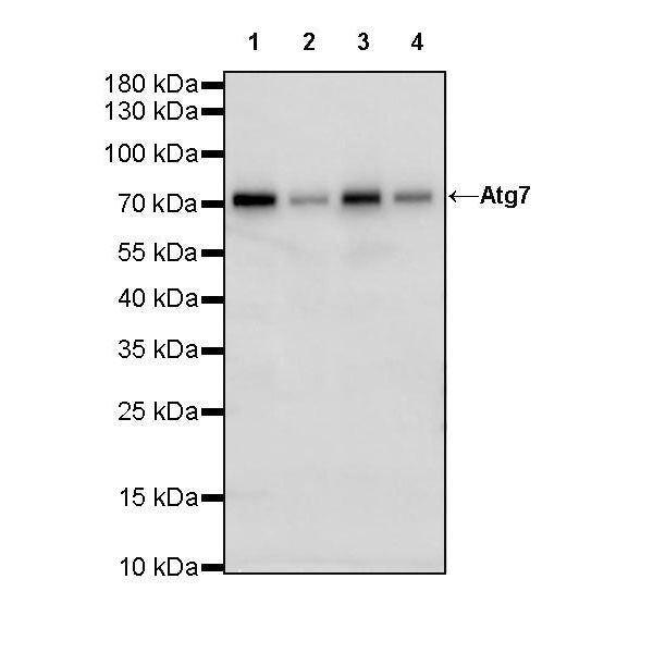
WB result of Atg7 Rabbit mAb
Primary antibody: Atg7 Rabbit mAb at 1/1000 dilution
Lane 1: Jurkat whole cell lysate 20 µg
Lane 2: HepG2 whole cell lysate 20 µg
Lane 3: HeLa whole cell lysate 20 µg
Lane 4: HT-29 whole cell lysate 20 µg
Negative control: Jurkat whole cell lysate
Secondary antibody: Goat Anti-Rabbit IgG, (H+L), HRP conjugated at 1/10000 dilution
Predicted MW: 77 kDa
Observed MW: 75 kDa
Atg7 Recombinant Rabbit mAb (S-R302)
Atg7 Recombinant Rabbit mAb (S-R302)
Price:
Regular price
$100 USD
Regular price
Sale price
$100 USD
Unit price
per
For shipping services or bulk orders, you may request a quotation.
Secure checkout with

View full details
Product Details
Product Details
Product Specification
| Host | Rabbit |
| Antigen | ATG7 |
| Synonyms | Ubiquitin-like modifier-activating enzyme ATG7, ATG12-activating enzyme E1 ATG7, Autophagy-related protein 7 (APG7-like; hAGP7), Ubiquitin-activating enzyme E1-like protein, APG7L |
| Location | Cytoplasm |
| Accession | O95352 |
| Clone Number | S-R302 |
| Antibody Type | Recombinant mAb |
| Isotype | IgG |
| Application | WB, IHC-P, IP |
| Reactivity | Hu |
| Purification | Protein A |
| Concentration | 0.5 mg/ml |
| Conjugation | Unconjugated |
| Physical Appearance | Liquid |
| Storage Buffer | PBS, 40% Glycerol, 0.05%BSA, 0.03% Proclin 300 |
| Stability & Storage | 12 months from date of receipt / reconstitution, -20 °C as supplied |
Dilution
| application | dilution | species |
| WB | 1:1000 | null |
| IHC-P | 1:500 | null |
| IP | 1:50 | null |
Background
ATG 7, present in both plant and animal genomes, acts as an essential protein for cell degradation and its recycling. The sequence associates with the ubiquitin- proteasome system, UPS, required for the unique development of an autophagosomal membrane and fusion within cells. During the initiation of autophagy, ATG7 acts like an E-1 enzyme for ubiquitin-like proteins (UBL) such as ATG12 and ATG8. ATG7 helps these UBL proteins in targeting their molecule by binding to them and activating their transfer to an E-2 enzyme. ATG7's role in both of these autophagy-specific UBL systems makes it an essential regulator of autophagosome assembly.
Picture
Picture
Western Blot
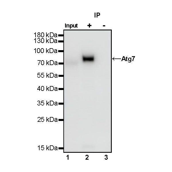
IP
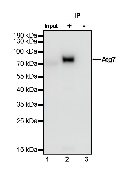
Atg7 Rabbit mAb at 1/50 dilution (1 µg) immunoprecipitating Atg7 in 0.4 mg Jurkat whole cell lysate.
Western blot was performed on the immunoprecipitate using Atg7 Rabbit mAb at 1/1000 dilution.
Secondary antibody (HRP) for IP was used at 1/400 dilution.
Lane 1: Jurkat whole cell lysate 20 µg (Input)
Lane 2: Atg7 Rabbit mAb IP in Jurkat whole cell lysate
Lane 3: Rabbit monoclonal IgG IP in Jurkat whole cell lysate
Predicted MW: 77 kDa
Observed MW: 75 kDa
Immunohistochemistry
IHC shows positive staining in paraffin-embedded human tonsil. Anti-Atg7 antibody was used at 1/500 dilution, followed by a HRP Polymer for Mouse & Rabbit IgG (ready to use). Counterstained with hematoxylin. Heat mediated antigen retrieval with Tris/EDTA buffer pH9.0 was performed before commencing with IHC staining protocol.
IHC shows positive staining in paraffin-embedded human breast cancer. Anti-Atg7 antibody was used at 1/500 dilution, followed by a HRP Polymer for Mouse & Rabbit IgG (ready to use). Counterstained with hematoxylin. Heat mediated antigen retrieval with Tris/EDTA buffer pH9.0 was performed before commencing with IHC staining protocol.
IHC shows positive staining in paraffin-embedded human cervical squamous cell carcinoma. Anti-Atg7 antibody was used at 1/500 dilution, followed by a HRP Polymer for Mouse & Rabbit IgG (ready to use). Counterstained with hematoxylin. Heat mediated antigen retrieval with Tris/EDTA buffer pH9.0 was performed before commencing with IHC staining protocol.
IHC shows positive staining in paraffin-embedded human pancreatic cancer. Anti-Atg7 antibody was used at 1/500 dilution, followed by a HRP Polymer for Mouse & Rabbit IgG (ready to use). Counterstained with hematoxylin. Heat mediated antigen retrieval with Tris/EDTA buffer pH9.0 was performed before commencing with IHC staining protocol.