UniOne® TR-FRET Human cAMP Hirange Detection Kit
UniOne® TR-FRET Human cAMP Hirange Detection Kit
Product Details
Product Details
Product Specification
| Host | Human |
| Stability & Storage | -80℃ |
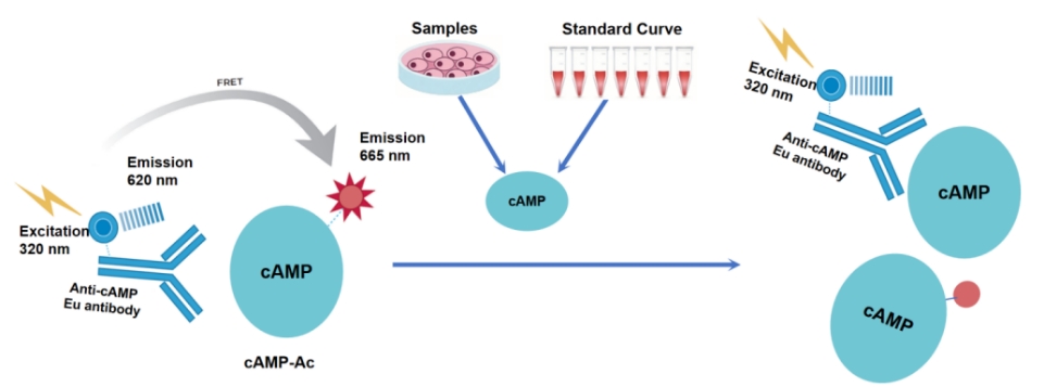
Background
The kit employs homogeneous time-resolved fluorescence (TR-FRET) technology, enabling precise measurement of cellular cAMP production. This method allows rapid detection of cAMP accumulation changes during the inhibition or activation of Gs or Gi signaling pathways.
As shown in the figure, the Eu-labeled anti-cAMP antibody (TR-FRET donor) and the Ac-labeled cAMP (TR-FRET acceptor) bind directly. When the donor antibody and acceptor antibody are in proximity, excitation of the donor antibody triggers fluorescence resonance energy transfer (FRET) to the acceptor antibody, resulting in a specific 665 nm emission signal. When cAMP from cell samples or standard curves is present, it competes with the Ac-labeled cAMP for binding to the Eu-labeled anti-cAMP antibody, thereby reducing the FRET signal. This specific signal is inversely proportional to the extent of interaction between the Eu-labeled anti-cAMP antibody and the Ac-labeled cAMP. The homogeneous assay is simple to perform and requires no washing steps.
Components
Component |
Concentration |
100T |
500T |
2500T |
10000T |
Storage Temperature |
cAMP calibrator |
4mM |
5μL |
5μL |
25μL |
100μL |
-80℃ |
Anti-cAMP Eu antibody |
100× |
5μL |
25μL |
125μL |
500μL |
-80℃ |
cAMP-Ac |
100× |
5μL |
25μL |
125μL |
500μL |
-80℃ |
Lysis & Detect Buffer |
1× |
2mL |
10mL |
50mL |
200mL |
-80℃ |
Stimulation Buffer (without IBMX) |
1× |
2mL |
10mL |
50mL |
200mL |
-80℃ |
Note: Aliquot immediately upon first thawing and store at the recommended temperature. Avoid storage after dilution and repeated freeze-thaw cycles.
Protocol
Here is the translated content in English, maintaining the original formatting:```html
1. Reagent Preparation
1.1 Thaw all reagents to room temperature before use. The reaction system for the 384-well plate is 20μL (reagent volumes are shown in the table below). Calculate the required volume before preparation. The following preparation is for reference only, using 500 reactions as an example.
Table 1. Reagent Preparation and Volumes
Reagent Name |
Preparation |
Volume per Well (μL) |
Anti-cAMP Eu antibody |
Take 25μL of Anti-cAMP Eu antibody stock, dilute to 2.5mL with Lysis & Detect Buffer, mix well for use. |
5μL |
cAMP-Ac |
Take 25μL of cAMP-Ac stock, dilute to 2.5mL with Lysis & Detect Buffer, mix well for use; |
5μL |
1.2 Gradient Dilution of Test Samples
Prepare according to experimental conditions. The following is for reference only.
Using cAMP calibrator as an example, the diluent is Stimulation Buffer. Adjust sample concentrations as needed.
Table 2. cAMP Calibrator Preparation Table
|
Preparation Concentration (nM) |
Final Concentration (nM) |
Preparation Method |
1 |
40,000.000 |
10,000.000 |
1μL of 4mM stock + 99μL Stimulation Buffer |
2 |
13,333.333 |
3,333.333 |
20μL of ① + 40μL Stimulation Buffer |
3 |
4,444.444极left;"> 1,111.111 |
20μL of ② + 40μL Stimulation Buffer |
|
4 |
1,481.481 |
370.370 |
20μL of ③ + 40μL Stimulation Buffer |
5 |
493.827 |
123.457 |
20μL of ④ + 40μL Stimulation Buffer |
6 |
164.609 |
41.152 |
20μL of ⑤ + 40μL Stimulation Buffer |
7 |
54.870 |
13.717 |
20μL of ⑥ + 40μL Stimulation Buffer |
8 |
18.290 |
4.572 |
20μL of ⑦ + 40μL Stimulation Buffer |
9 |
6.097 |
1.524 |
20μL of ⑧ + 40μL Stimulation Buffer |
10 |
2.032 |
0.508 |
20μL of ⑨ + 40μL Stimulation Buffer |
11 |
0.677 |
0.169 |
20μL of ⑩ + 40μL Stimulation Buffer |
⑫ |
0.226 |
0.056 |
20μL of ⑪ + 40μL Stimulation Buffer |
Blank |
0 |
0 |
40μL of 1× Stimulation Buffer |
1.3 Pre-Treatment for Cell-Based Experiments
①Stimulation Buffer is used for diluting cells and stimulating drugs. The Stimulation Buffer provided in the kit does not contain IBMX. Add an appropriate concentration of IBMX to prevent cAMP degradation, depending on experimental objectives.
②Before formal experiments, optimize the number of cells per well and the incubation time with drugs based on cell and drug characteristics to ensure cAMP levels fall within the linear range of the calibration curve.
2. Sample Loading and Controls
2.1 cAMP Standard Curve: 5μL of gradient-diluted cAMP reference, 5μL Stimulation Buffer, 5μL Anti-cAMP Eu antibody working solution. Incubate for 10min, then add 5μL cAMP-Ac.
2.2 Test Samples: 5μL of gradient-diluted sample, 5μL Stimulation Buffer, 5μL Anti-cAMP Eu antibody working solution. Incubate for 10min, then add 5μL cAMP-Ac.
2.3 Cell-Based Experiments: Based on pre-experiment results, add 5μL cells and 5μL gradient-diluted drug per well. After incubation (optimized time), add 5μL Anti-cAMP Eu antibody working solution, incubate at room temperature for 10min, then add 5μL cAMP-Ac.
2.4 Negative Control (NC): 5μL Stimulation Buffer instead of sample or 5μL untreated cells.
|
cAMP Standard Curve |
Test Samples |
Cell Experiments |
Negative Control (NC) |
|
Step 1
|
5μL cAMP calibrator |
5μL test sample |
5μL cells |
5μL Stimulation Buffer or 5μL cells |
5μL Stimulation Buffer |
5μL Stimulation Buffer极left;"> 5μL drug※ |
5μL Stimulation Buffer |
||
5μL Anti-cAMP Eu antibody working solution | ||||
Incubate at room temperature for 10min | ||||
Step 2 |
5μL cAMP-Ac |
|||
|
Seal plate, centrifuge at 1000rpm for 1min to mix, incubate at room temperature for 120min, then read plate. | ||||
•Optimize incubation time for cells and drugs experimentally. Add subsequent reagents after drug stimulation.
3.Detection
Measure using a TR-FRET-compatible microplate reader. Excitation at 320/340nm, emission at 620nm and 665nm.
【Data Analysis】
1) Calculate signal ratio (Ratio): (665nm signal / 620nm signal) × 10000.
Ratio = (665/620) × 10000
2) Calculate CV (%):
CV (%) = (Standard Deviation / Mean Ratio) × 100%
【Example Data】
The following data is for illustration only and may vary depending on the reader.

Note: *Example data only. Recommended plate: 384-well, white, shallow-well.
```