WB result of PEG10 Rabbit mAb Primary antibody: PEG10 Rabbit mAb at 1/1000 dilution Lane 1: 293T whole cell lysate 20 µg Lane 2: HeLa whole cell lysate 20 µg Lane 3: HepG2 whole cell lysate 20 µg Negative control: 293T whole cell lysate Secondary antibody: Goat Anti-Rabbit IgG, (H+L), HRP conjugated at 1/10000 dilution Predicted MW: 80 kDa Observed MW: 110 kDa
PEG10 Recombinant Rabbit mAb (SDT-442-60)
PEG10 Recombinant Rabbit mAb (SDT-442-60)
Price:
Regular price
$100 USD
Regular price
Sale price
$100 USD
Unit price
per
For shipping services or bulk orders, you may request a quotation.
Secure checkout with

View full details
Product Details
Product Details
Product Specification
| Host | Rabbit |
| Antigen | PEG10 |
| Synonyms | Retrotransposon-derived protein PEG10, Embryonal carcinoma differentiation-regulated protein, Mammalian retrotransposon-derived protein 2, MEF3-like protein 1, Paternally expressed gene 10 protein, Ty3/Gypsy-like protein, MEF3L1 |
| Immunogen | Recombinant Protein |
| Location | Cytoplasm |
| Accession | Q86TG7 |
| Clone Number | SDT-442-60 |
| Antibody Type | Recombinant mAb |
| Application | WB, IHC-P, ICC |
| Reactivity | Hu |
| Purification | Protein A |
| Concentration | 0.5 mg/ml |
| Conjugation | Unconjugated |
| Physical Appearance | Liquid |
| Storage Buffer | PBS, 40% Glycerol, 0.05% BSA, 0.03% Proclin 300 |
| Stability & Storage | 12 months from date of receipt / reconstitution, -20 °C as supplied |
Dilution
| application | dilution | species |
| WB | 1:1000 | null |
| ICC | 1:500 | null |
| IHC-P | 1:1000 | null |
Background
Retrotransposon-derived protein PEG10 is a protein that in humans is encoded by the PEG10 gene. Binds its own mRNA in the 5'-UTR region, in the region near the boundary between the nucleocapsid (NC) and protease (PRO) coding sequences and in the beginning of the 3'-UTR region [PubMed:34413232].
Picture
Picture
Western Blot
Immunohistochemistry
IHC shows positive staining in paraffin-embedded human breast carcinoma. Anti-PEG10 antibody was used at 1/1000 dilution, followed by a HRP Polymer for Mouse & Rabbit IgG (ready to use). Counterstained with hematoxylin. Heat mediated antigen retrieval with Tris/EDTA buffer pH9.0 was performed before commencing with IHC staining protocol.
Negative control: IHC shows negative staining in paraffin-embedded human breast. Anti-PEG10 antibody was used at 1/1000 dilution, followed by a HRP Polymer for Mouse & Rabbit IgG (ready to use). Counterstained with hematoxylin. Heat mediated antigen retrieval with Tris/EDTA buffer pH9.0 was performed before commencing with IHC staining protocol.
IHC shows positive staining in paraffin-embedded human hepatocellular carcinoma. Anti-PEG10 antibody was used at 1/1000 dilution, followed by a HRP Polymer for Mouse & Rabbit IgG (ready to use). Counterstained with hematoxylin. Heat mediated antigen retrieval with Tris/EDTA buffer pH9.0 was performed before commencing with IHC staining protocol.
Negative control: IHC shows negative staining in paraffin-embedded human liver. Anti-PEG10 antibody was used at 1/1000 dilution, followed by a HRP Polymer for Mouse & Rabbit IgG (ready to use). Counterstained with hematoxylin. Heat mediated antigen retrieval with Tris/EDTA buffer pH9.0 was performed before commencing with IHC staining protocol.
IHC shows positive staining in paraffin-embedded human placenta. Anti-PEG10 antibody was used at 1/1000 dilution, followed by a HRP Polymer for Mouse & Rabbit IgG (ready to use). Counterstained with hematoxylin. Heat mediated antigen retrieval with Tris/EDTA buffer pH9.0 was performed before commencing with IHC staining protocol.
Negative control: IHC shows negative staining in paraffin-embedded human prostate. Anti-PEG10 antibody was used at 1/1000 dilution, followed by a HRP Polymer for Mouse & Rabbit IgG (ready to use). Counterstained with hematoxylin. Heat mediated antigen retrieval with Tris/EDTA buffer pH9.0 was performed before commencing with IHC staining protocol.
Immunocytochemistry
ICC shows positive staining in HepG2 cells. Anti-PEG10 antibody was used at 1/500 dilution (Green) and incubated overnight at 4°C. Goat polyclonal Antibody to Rabbit IgG - H&L (Alexa Fluor® 488) was used as secondary antibody at 1/1000 dilution. The cells were fixed with 4% PFA and permeabilized with 0.1% PBS-Triton X-100. Nuclei were counterstained with DAPI (Blue).
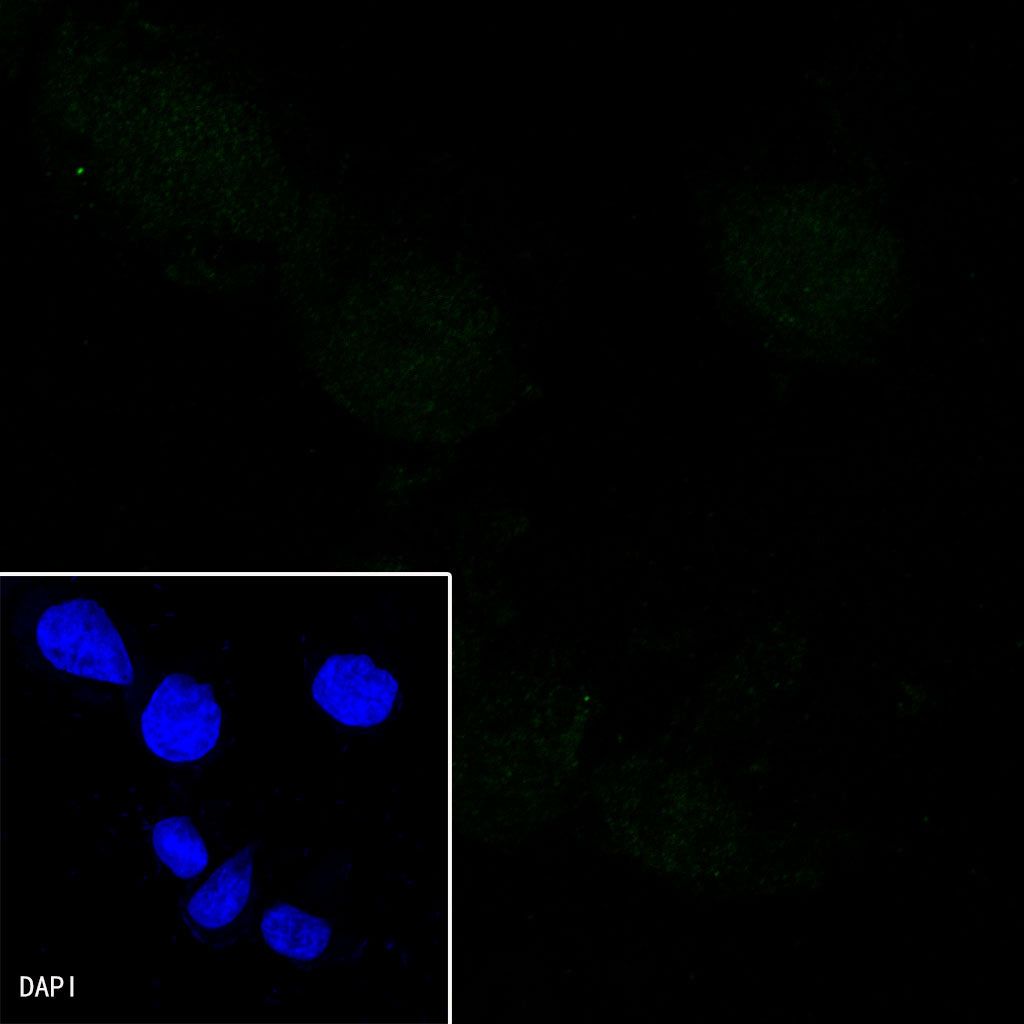
Negative control: ICC shows negative staining in 293T cells. Anti-PEG10 antibody was used at 1/500 dilution and incubated overnight at 4°C. Goat polyclonal Antibody to Rabbit IgG - H&L (Alexa Fluor® 488) was used as secondary antibody at 1/1000 dilution. The cells were fixed with 4% PFA and permeabilized with 0.1% PBS-Triton X-100. Nuclei were counterstained with DAPI (Blue).
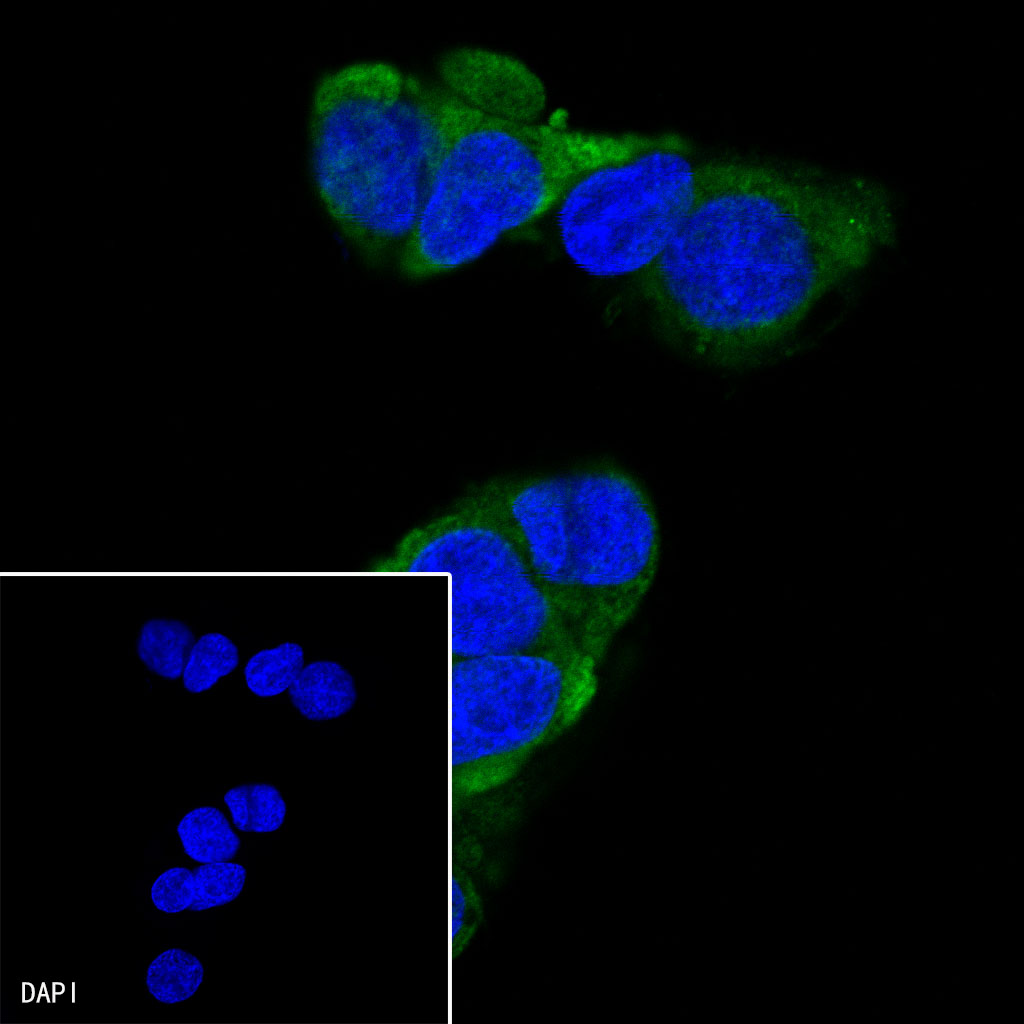
ICC shows positive staining in HepG2 cells. Anti-PEG10 antibody was used at 1/500 dilution (Green) and incubated overnight at 4°C. Goat polyclonal Antibody to Rabbit IgG - H&L (Alexa Fluor® 488) was used as secondary antibody at 1/1000 dilution. The cells were fixed with 4%PFA and permeabilized with 0.1% PBS-Triton X-100. Nuclei were counterstained with DAPI (Blue).
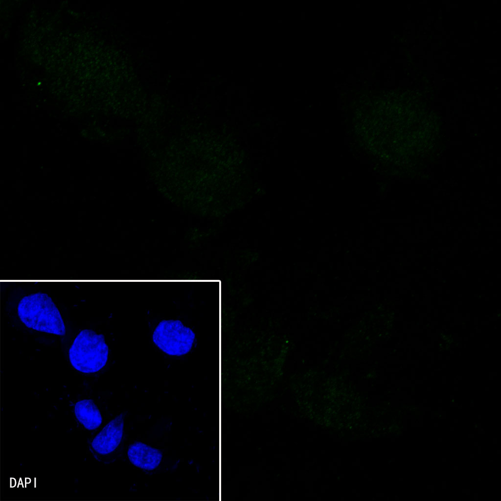
Negative control: ICC shows negative staining in 293T cells. Anti-PEG10 antibody was used at 1/500 dilution and incubated overnight at 4°C. Goat polyclonal Antibody to Rabbit IgG - H&L (Alexa Fluor® 488) was used as secondary antibody at 1/1000 dilution. The cells were fixed with 4%PFA and permeabilized with 0.1% PBS-Triton X-100. Nuclei were counterstained with DAPI (Blue).