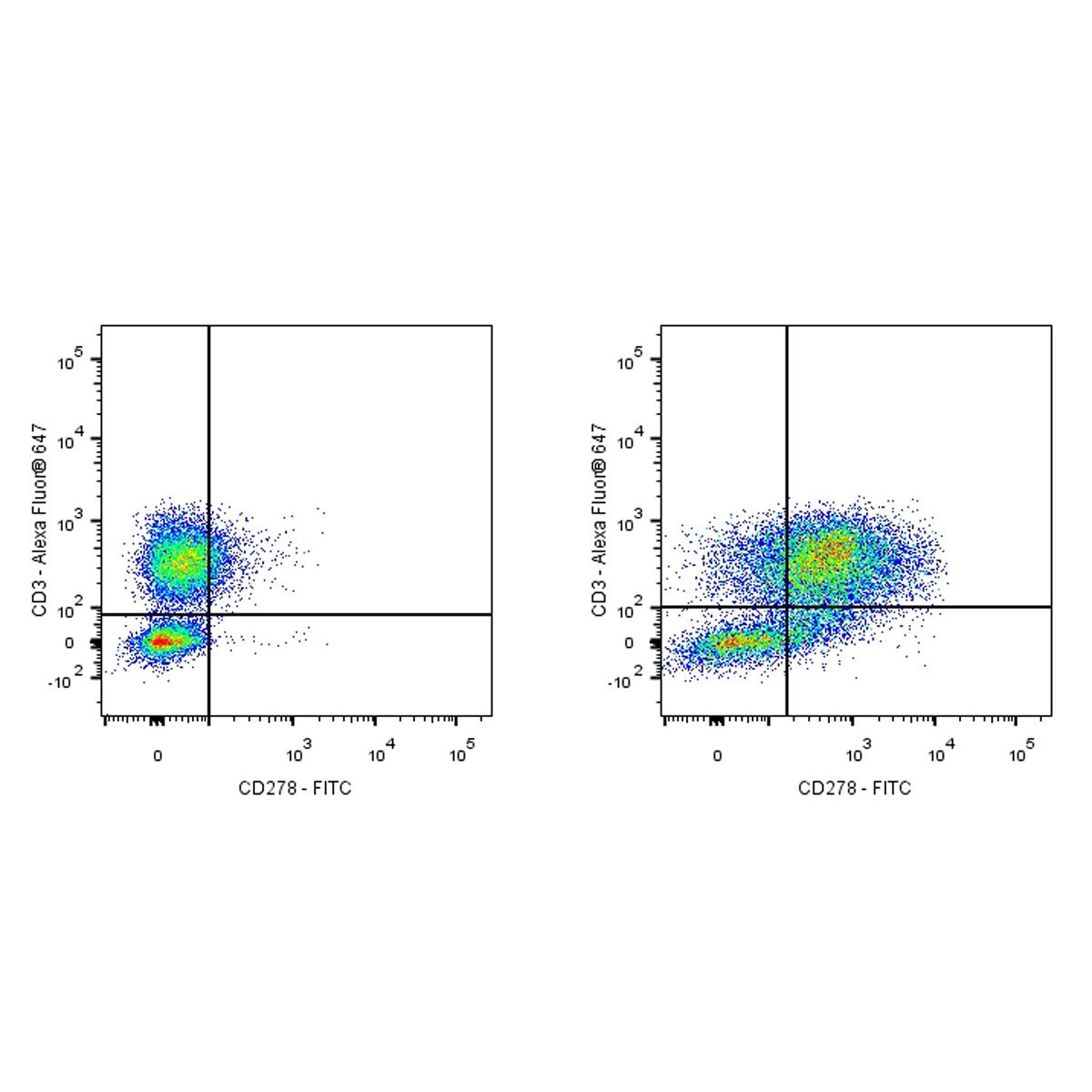
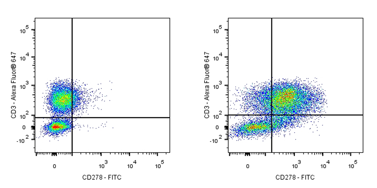
Treated 48h with 10μg/ml PHA human PBMC (human peripheral blood mononuclear cell) (Right panel) or untreated (Left panel) was stained with Alexa Fluor® 647 Mouse Anti-Human CD3 and SDT FITC Armenian Hamster Anti-Human CD278 Antibody at 5μl/test. Flow cytometry and data analysis were performed using BD FACSymphony™ A1 and FlowJo™ software.
FITC Armenian hamster Anti-Human/Mouse/Rat CD278 Antibody (S-R578)
FITC Armenian hamster Anti-Human/Mouse/Rat CD278 Antibody (S-R578)
Price:
Regular price
$110 USD
Regular price
Sale price
$110 USD
Unit price
per
For shipping services or bulk orders, you may request a quotation.
Secure checkout with

View full details
Product Details
Product Details
Product Specification
| Host | Armenian hamster |
| Antigen | CD278 |
| Synonyms | Inducible T-cell costimulatory; Activation-inducible lymphocyte immunomediatory molecule; ICOS; AILIM |
| Location | Cell membrane |
| Accession | Q9Y6W8 |
| Clone Number | S-R578 |
| Antibody Type | Recombinant mAb |
| Isotype | IgG |
| Application | FCM |
| Reactivity | Hu, Ms, Rt |
| Purification | Protein G |
| Concentration | 0.05mg/ml |
| Conjugation | FITC |
| Physical Appearance | Liquid |
| Storage Buffer | PBS, 25% Glycerol, 1% BSA, 0.3% Proclin 300 |
| Stability & Storage | 12 months from date of receipt / reconstitution, 2 to 8 °C as supplied. |
Dilution
| application | dilution | species |
| FCM | 5μl per million cells in 100μl volume | Hu |
Background
CD278, also known as ICOS (inducible T-cell co-stimulator), is a member of the CD28 superfamily and is expressed on activated T cells, including regulatory T cells (Tregs) and follicular helper T cells (Tfh). It functions as a homodimer and plays a crucial role in T cell activation, proliferation, and differentiation. ICOS binds to its ligand ICOSL (CD275), which is expressed on antigen-presenting cells (APCs) such as dendritic cells, B cells, and macrophages. This interaction enhances T cell-mediated immune responses, promotes cytokine production (e.g., IL-4, IL-13), and supports the survival and function of Tfh cells, which are critical for B cell antibody production. ICOS signaling is also involved in Th2 and Th17 cell differentiation and has been implicated in autoimmune and inflammatory diseases. In cancer immunotherapy, ICOS agonist antibodies have shown potential for enhancing anti-tumor immune responses when combined with other immunotherapies.
Picture
Picture
FC
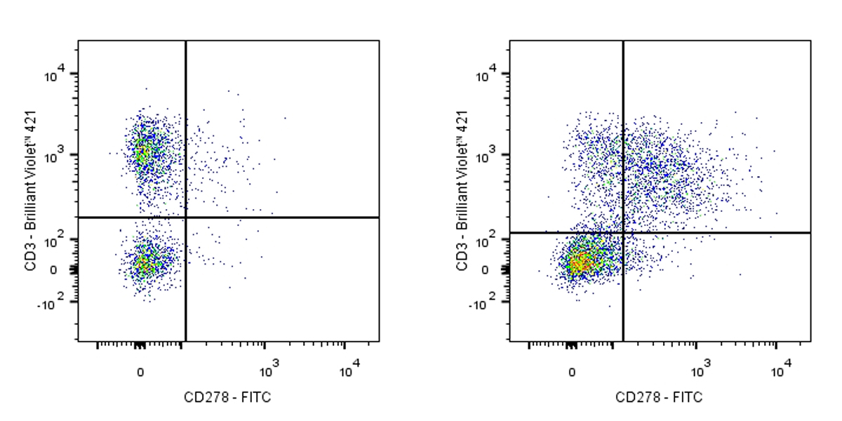
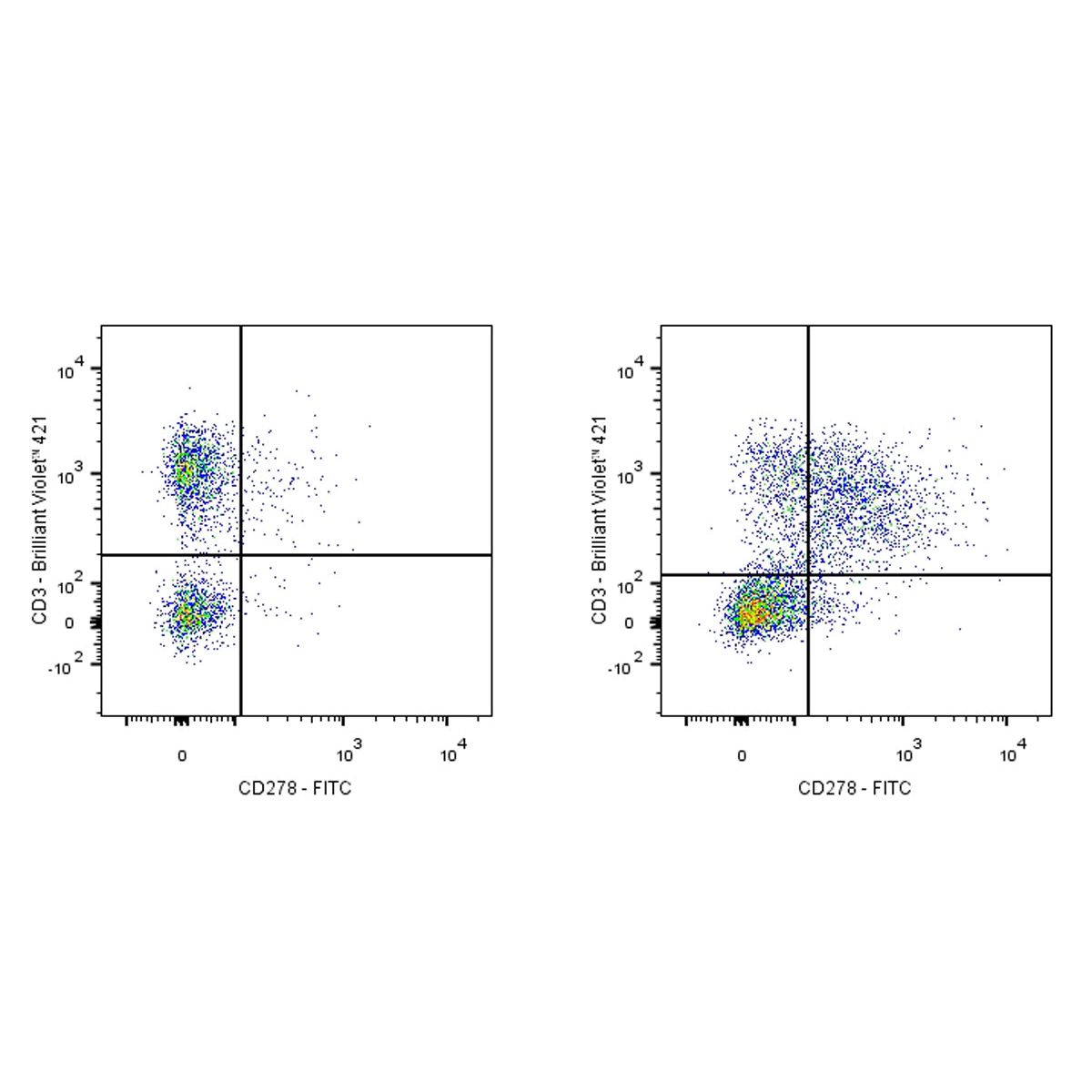
Treated overnight with 3μg/ml Concanavalin A C57BL/6 mouse splenocytes (Right panel) or untreated (Left panel) was stained with Alexa Fluor® 647 Rat Anti-Mouse CD3 and SDT FITC Armenian Hamster Anti-Human CD278 Antibody at 5μl/test. Flow cytometry and data analysis were performed using BD FACSymphony™ A1 and FlowJo™ software.