Product Specification
| Host |
Rabbit |
| Antigen |
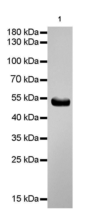
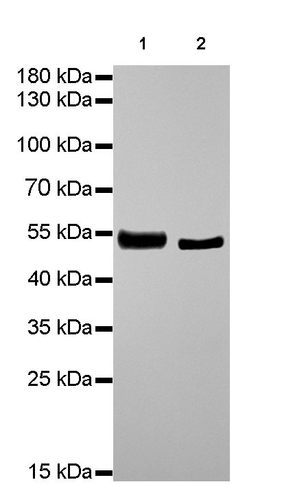
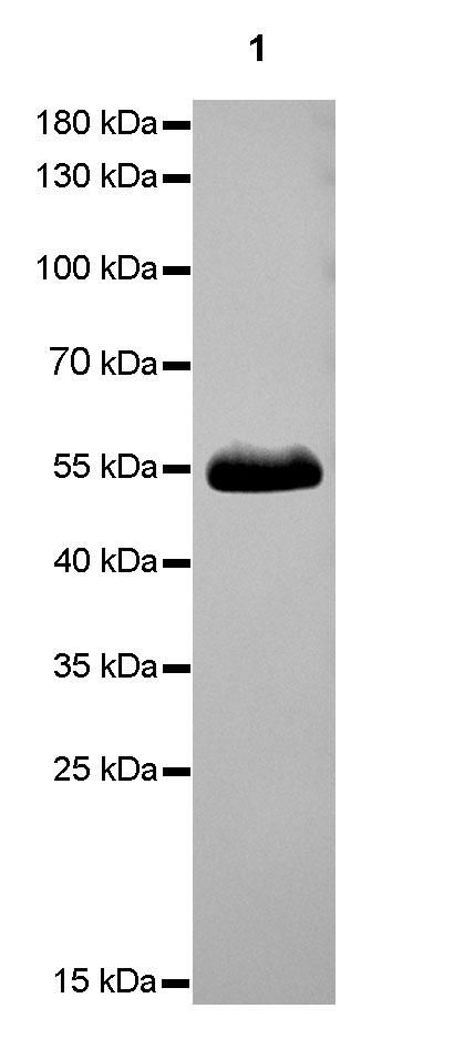
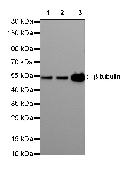
β-tubulin |
| Synonyms |
TUBB; TUBB5; Tubulin beta-5 chain |
| Immunogen |
Recombinant Protein |
| Location |
Intracellular |
| Accession |
P07437 |
| Clone Number |
SDT-R014 |
| Antibody Type |
Rabbit mAb |
| Application |
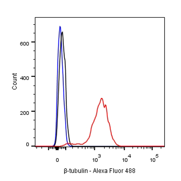
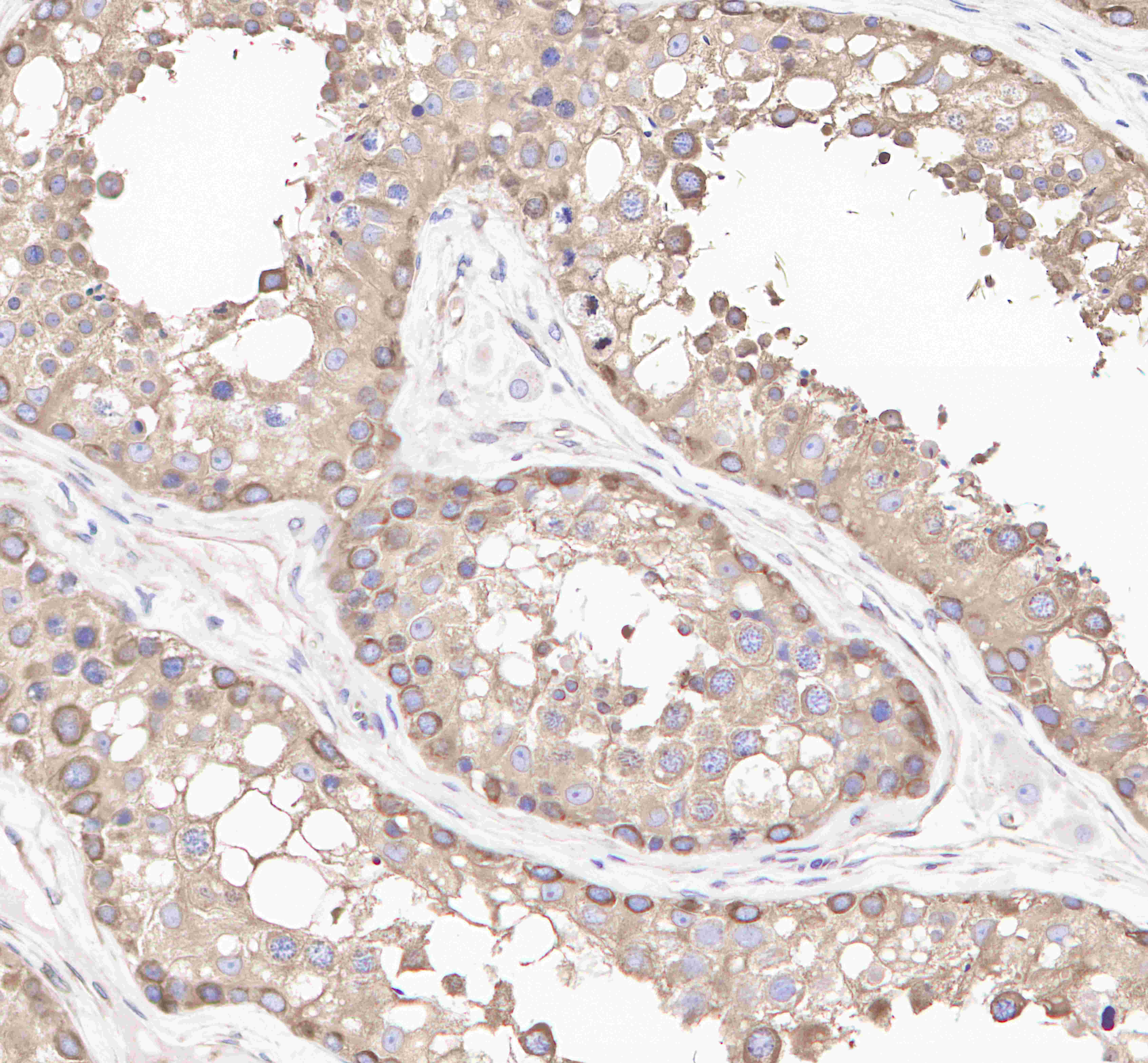
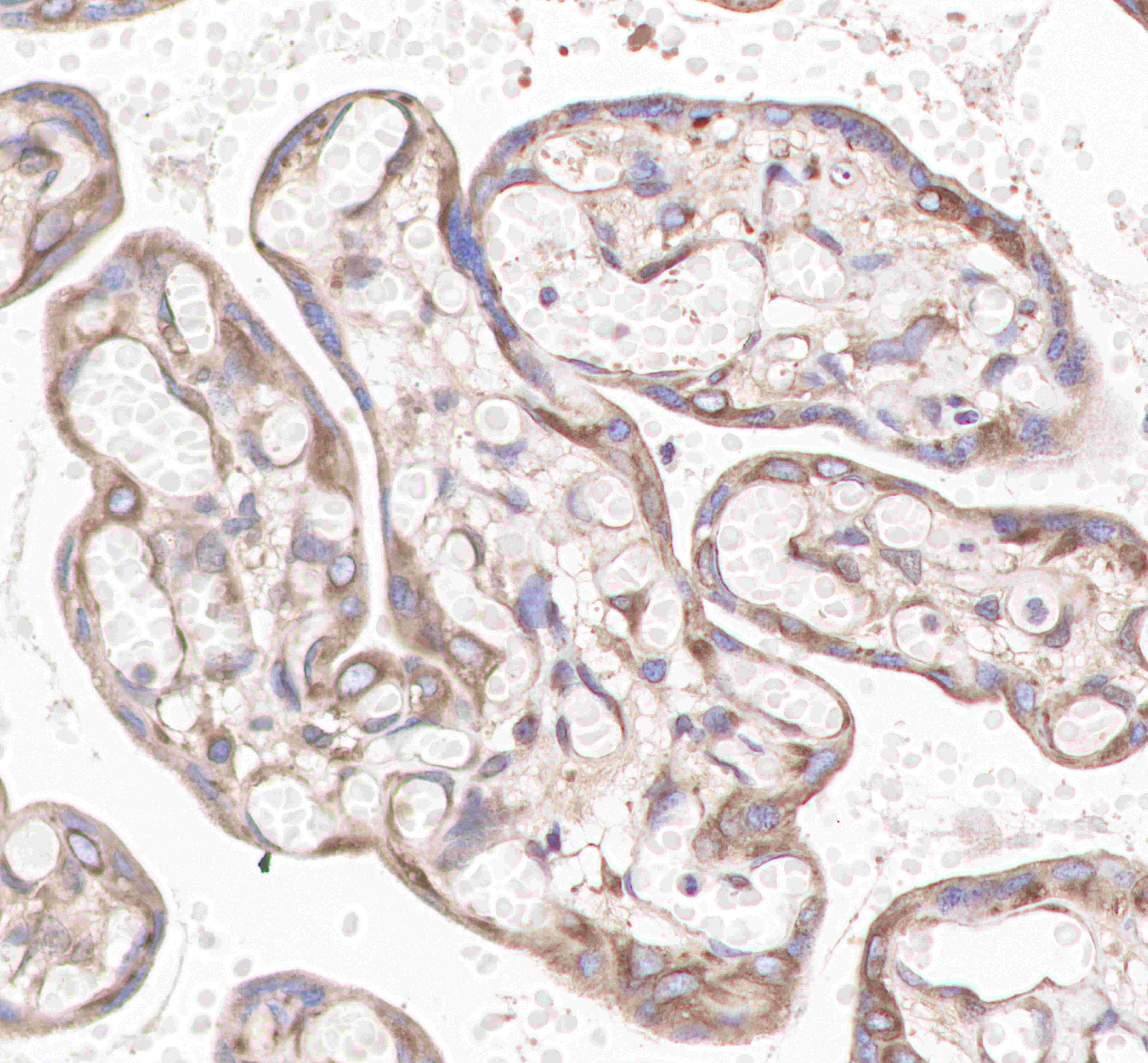
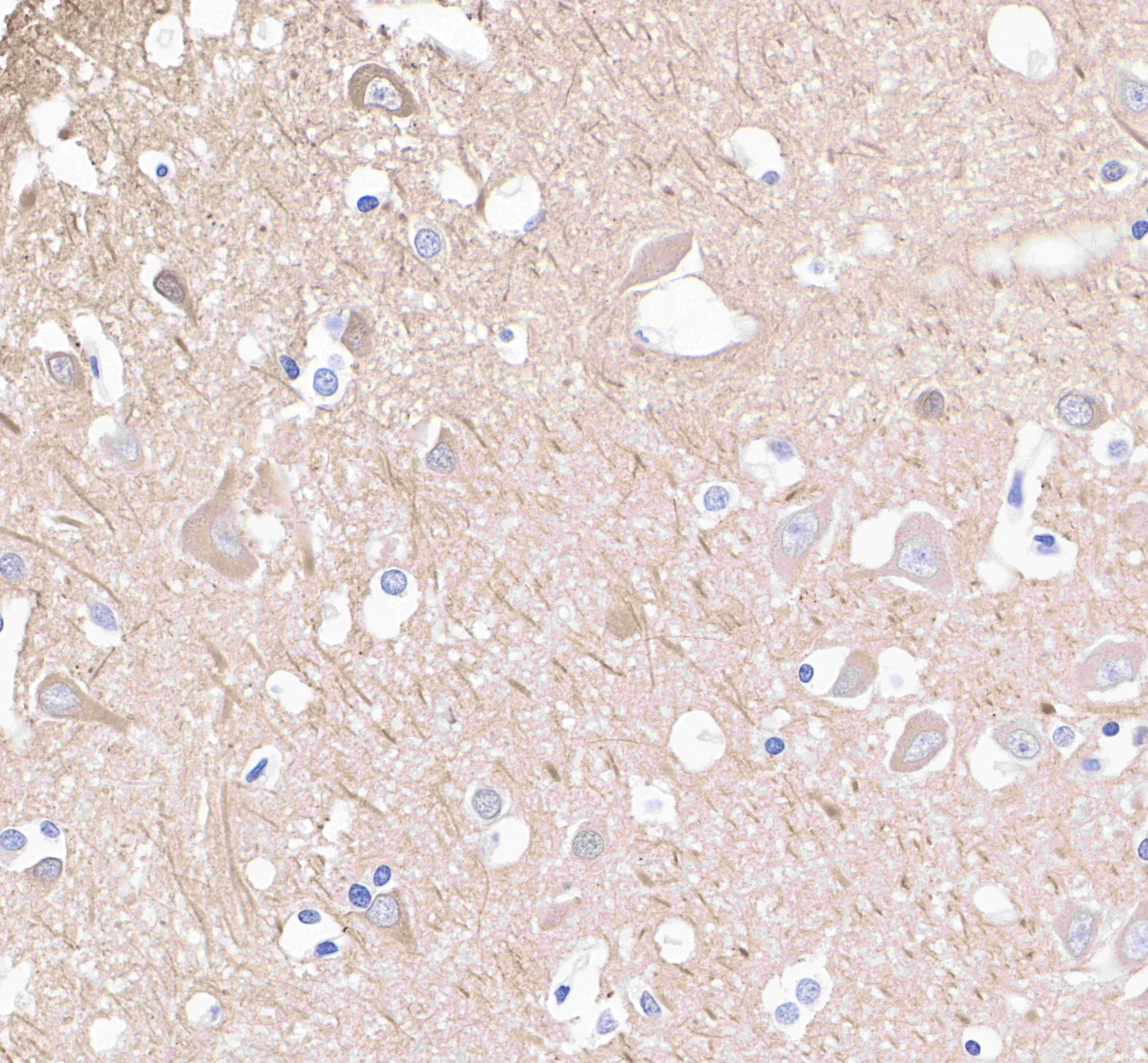
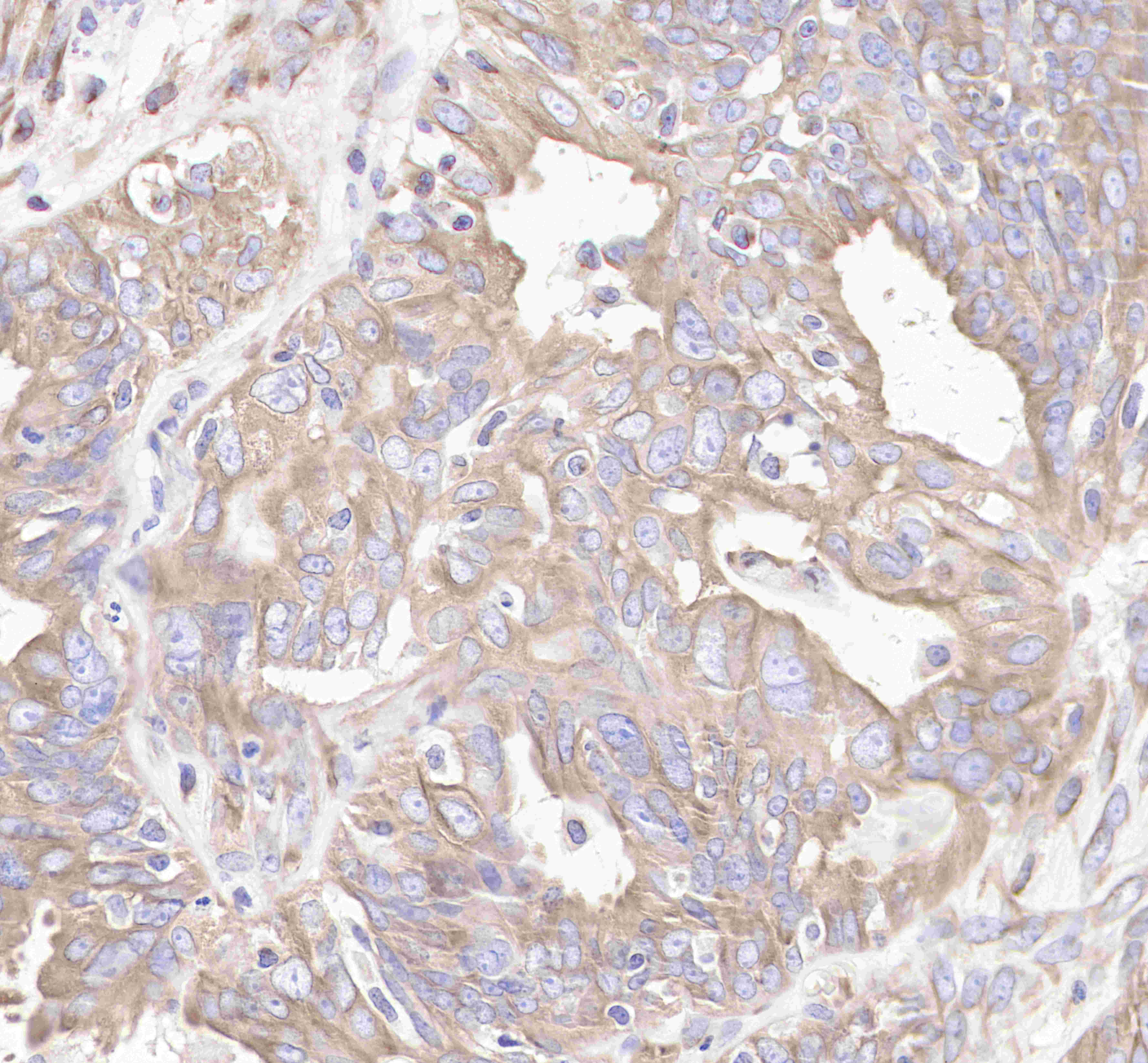
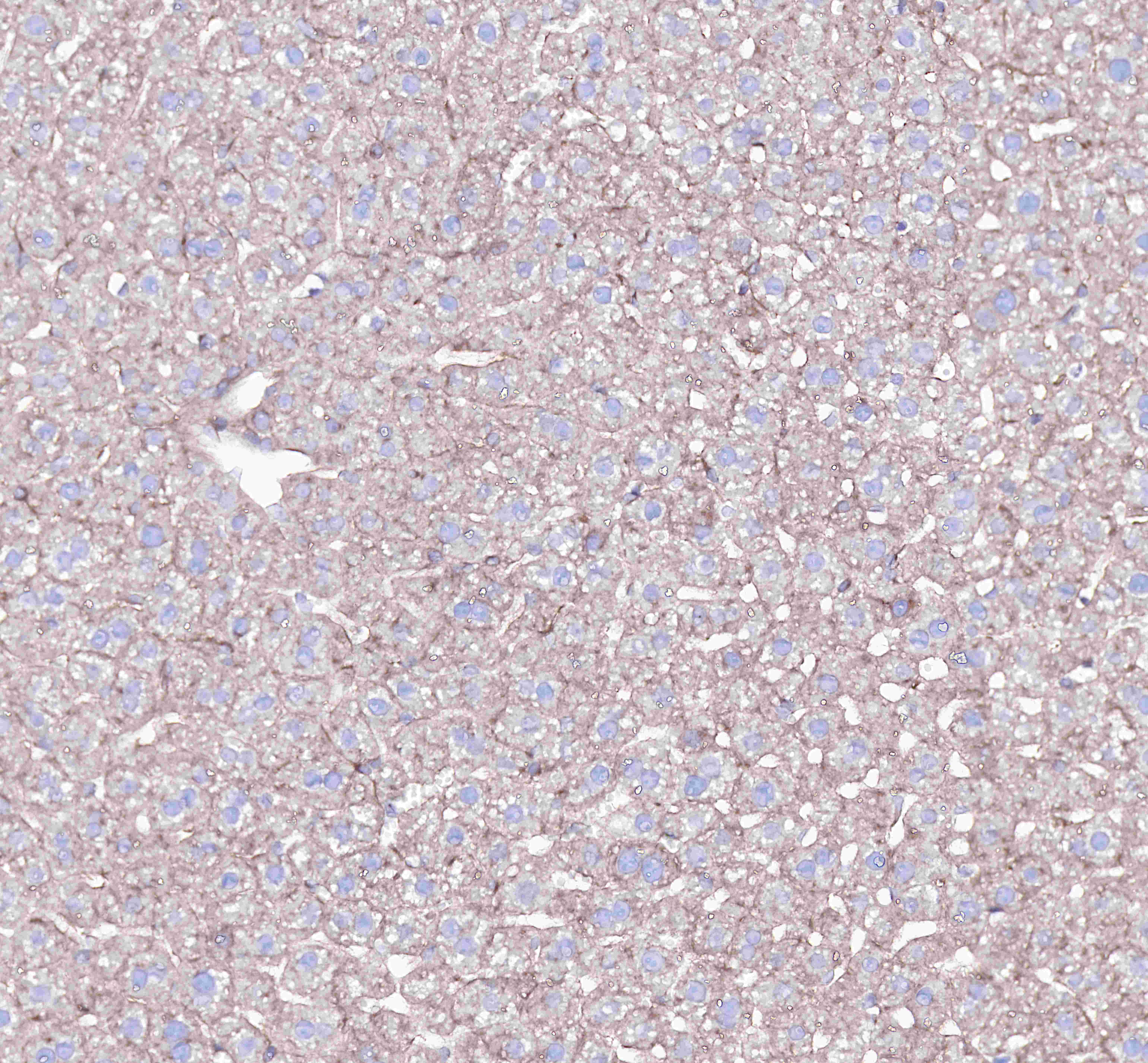
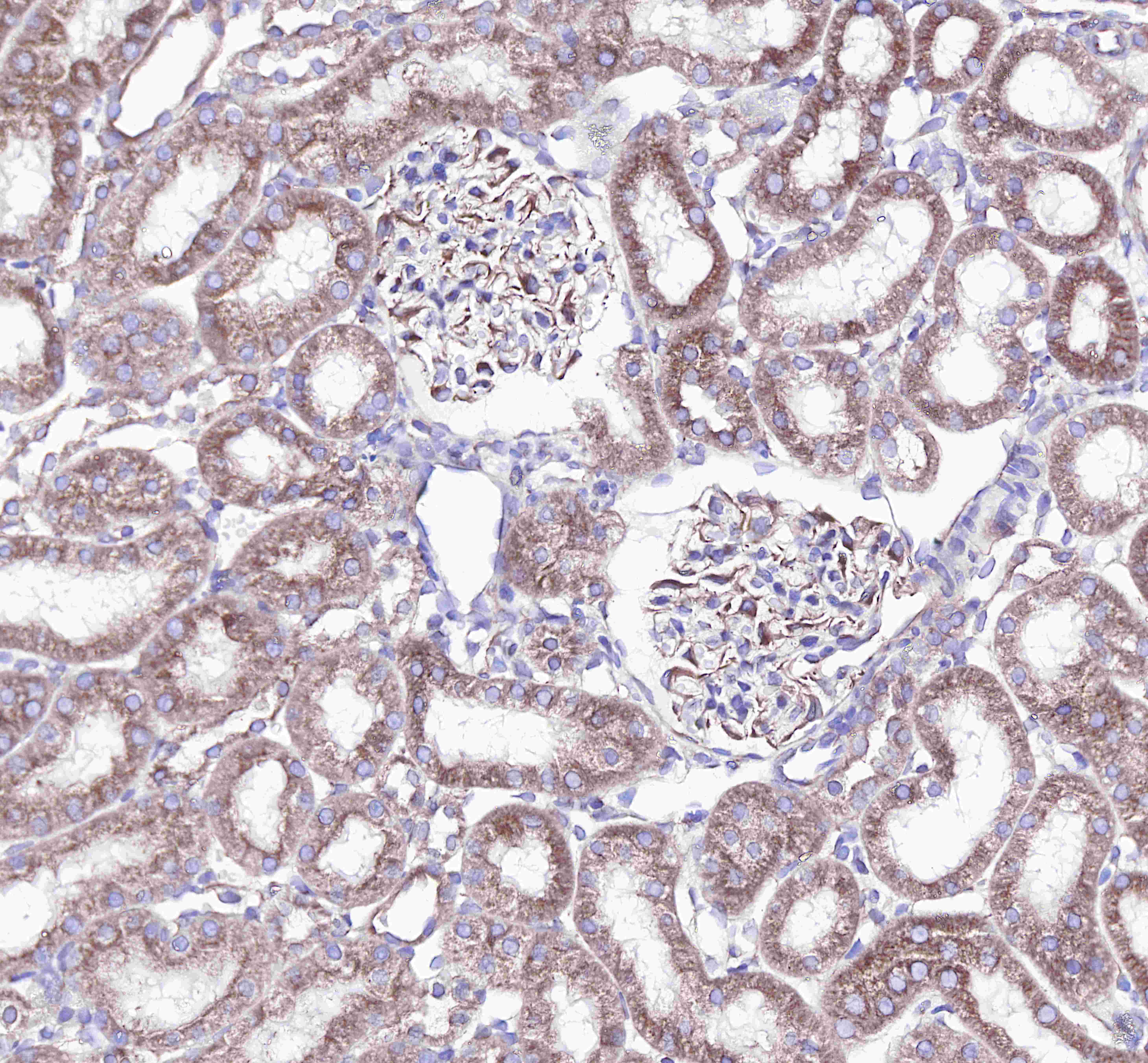
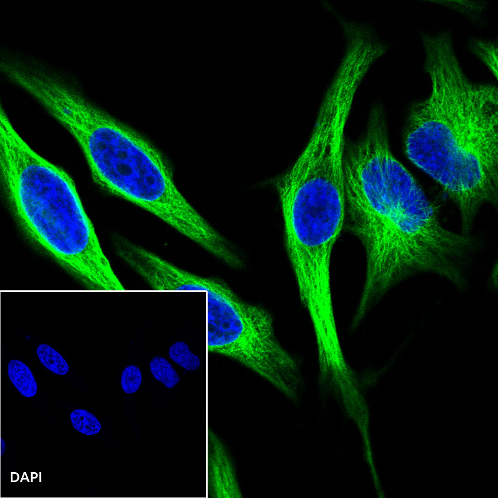
WB, IHC-P, ICC, ICFCM |
| Reactivity |
Hu, Ms, Rt |
| Purification |
Protein A |
| Concentration |
0.5mg/ml |
| Molecular Weight |
55kDa |
| Conjugation |
Unconjugated |
| Physical Appearance |
Liquid |
| Storage Buffer |
PBS, 40% Glycerol, 0.05%BSA, 0.03% Proclin 300 |
| Stability & Storage |
12 months from date of receipt / reconstitution, -20 °C as supplied. |
Dilution
| application |
dilution |
species |
| ICFCM |
1:500 |
|
| WB |
1:1000-1:10000 |
|
| IHC-P |
1:1000 |
|
| ICC |
1:500 |
|
Background
α- and β-tubulin polymerize into dynamic microtubules. In eukaryotes, microtubules are one of the major components of the cytoskeleton, and function in many processes, including structural support, intracellular transport, and DNA segregation. To form microtubules, the dimers of α- and β-tubulin bind to GTP and assemble onto the (+) ends of microtubules while in the GTP-bound state. The β-tubulin subunit is exposed on the plus end of the microtubule, while the α-tubulin subunit is exposed on the minus end. After the dimer is incorporated into the microtubule, the molecule of GTP bound to the β-tubulin subunit eventually hydrolyzes into GDP through inter-dimer contacts along the microtubule protofilament.