Product Specification
| Host |
Rabbit |
| Antigen |
MCM2 |
| Synonyms |
BM28, CCNL1, CDCL1, Minichromosome maintenance |
| Immunogen |
Synthetic Peptide |
| Location |
Nucleus |
| Accession |
P49736 |
| Clone Number |
SDT-018-68 |
| Antibody Type |
Rabbit mAb |
| Isotype |
IgG |
| Application |
WB, IHC-P, ICC, IF, ICFCM |
| Reactivity |
Hu, Ms |
| Purification |
Protein A |
| Research Area |
Epigenetics |
| Concentration |
0.5mg/ml |
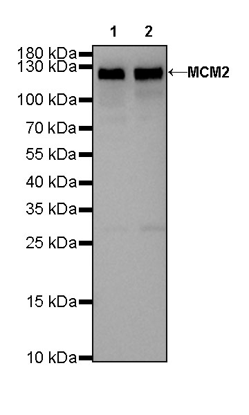
| Molecular Weight |
102kDa |
| Conjugation |
Unconjugated |
| Physical Appearance |
Liquid |
| Storage Buffer |
PBS, 40% Glycerol, 0.05%BSA, 0.03% Proclin 300 |
| Stability & Storage |
12 months from date of receipt / reconstitution, -20 °C as supplied |
Dilution
| application |
dilution |
species |
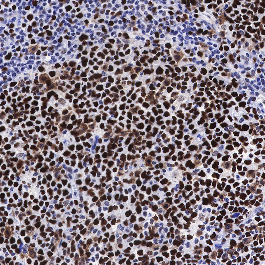
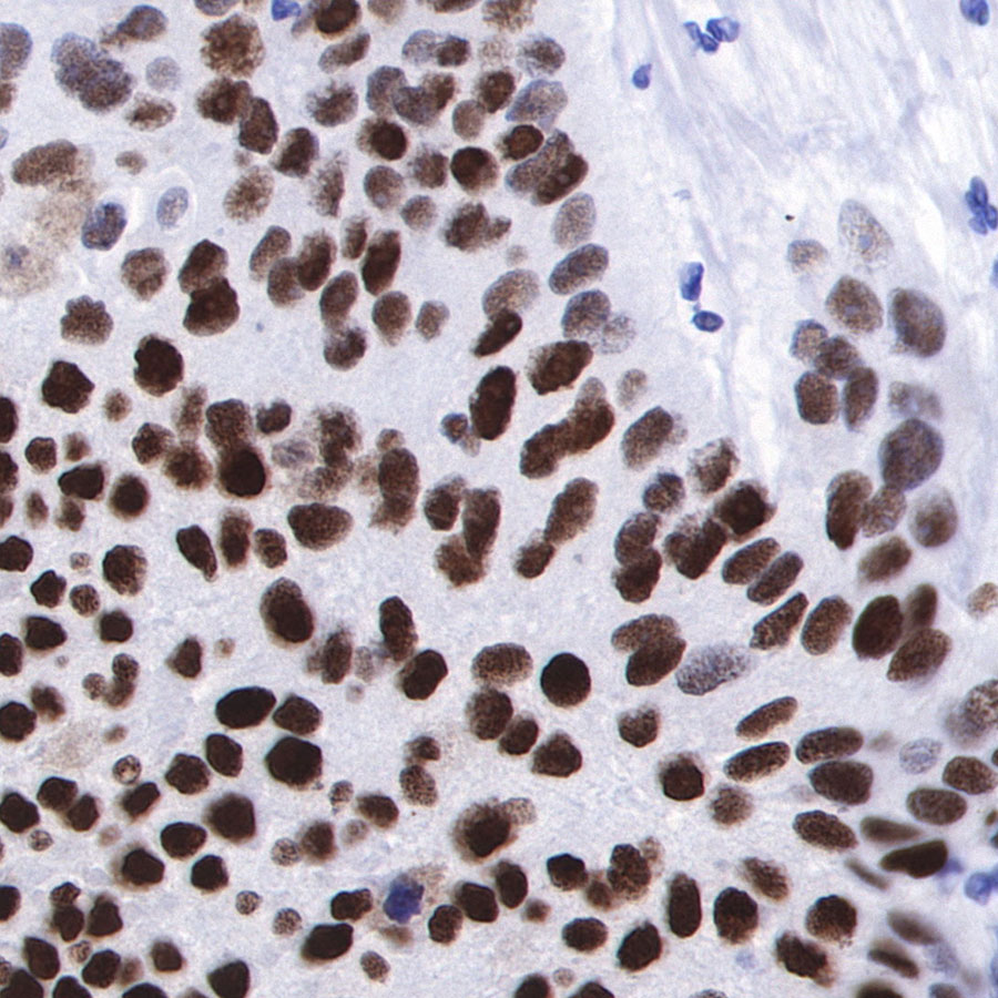
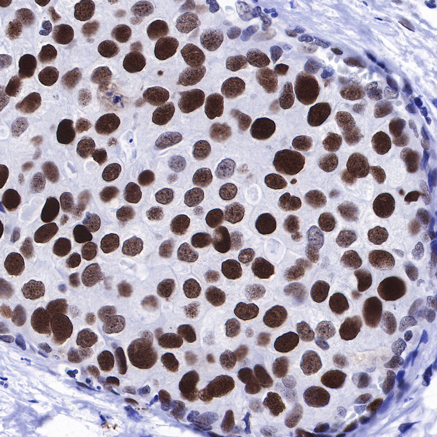
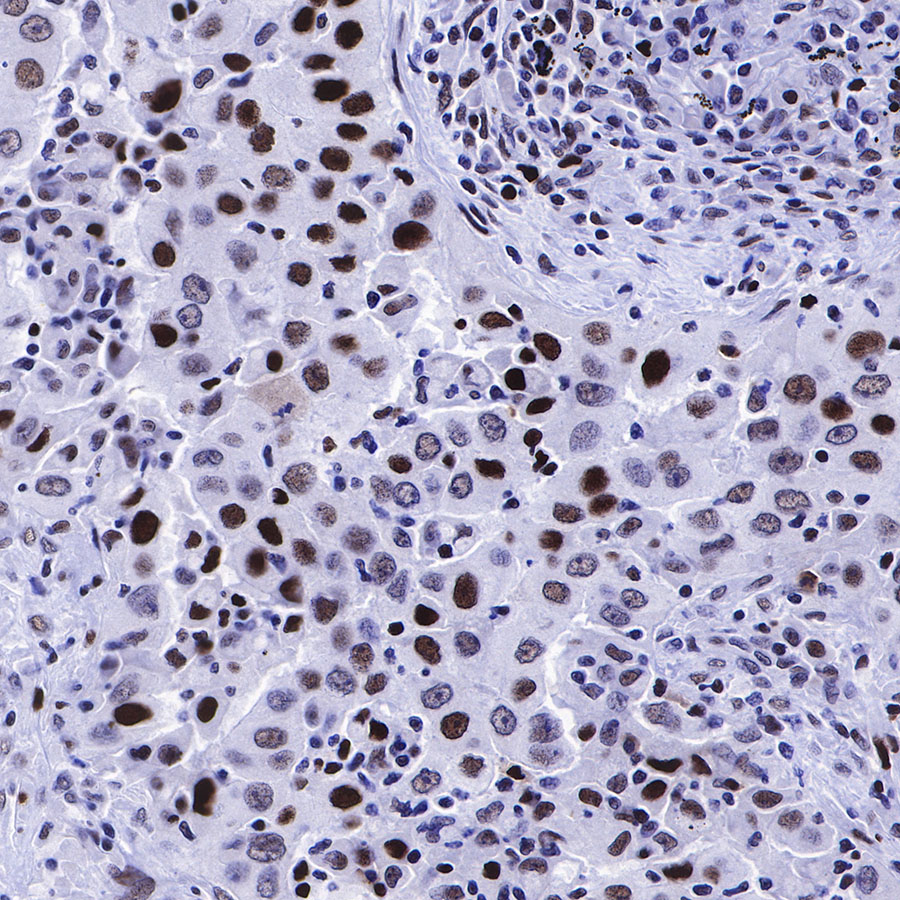
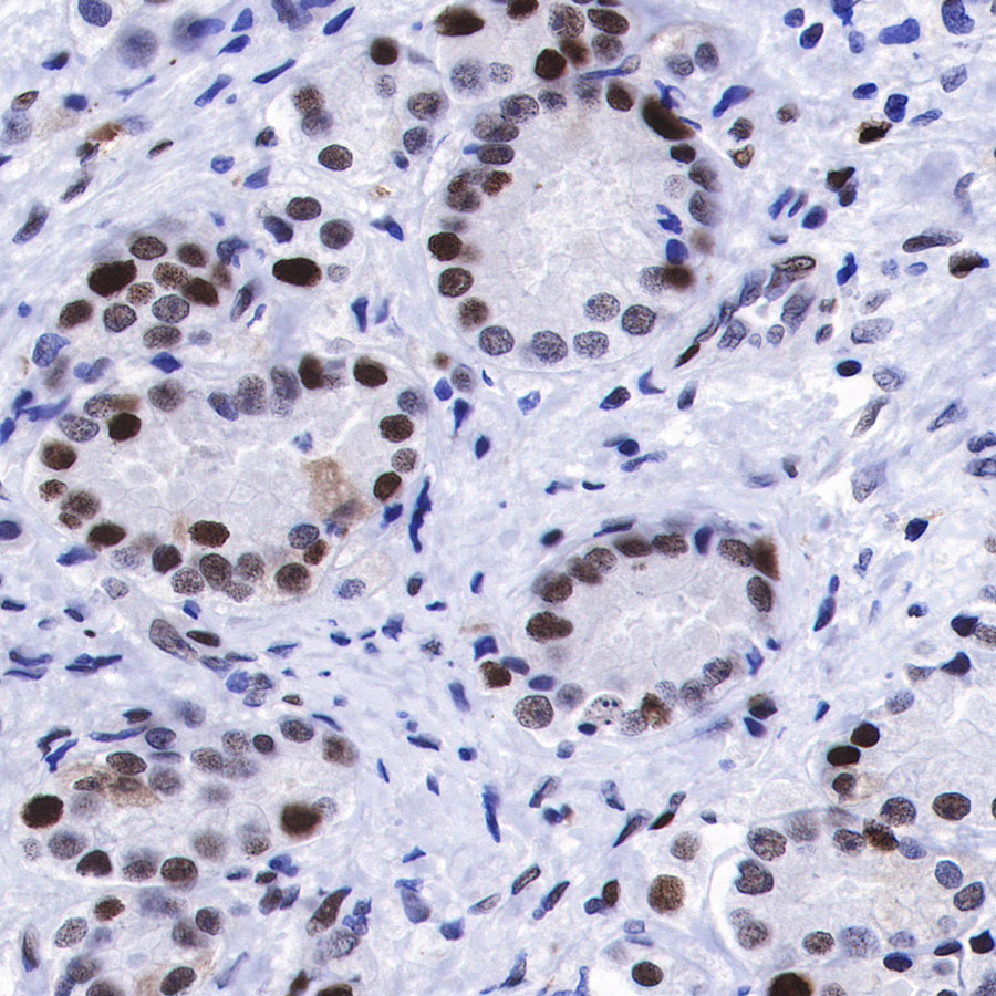
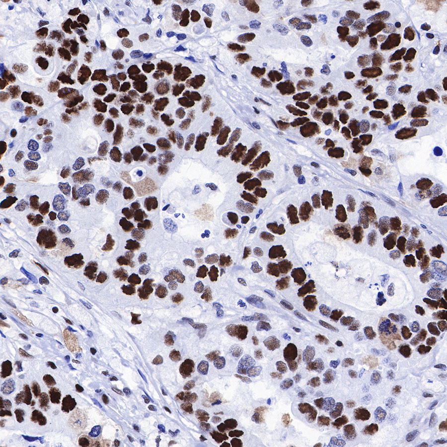
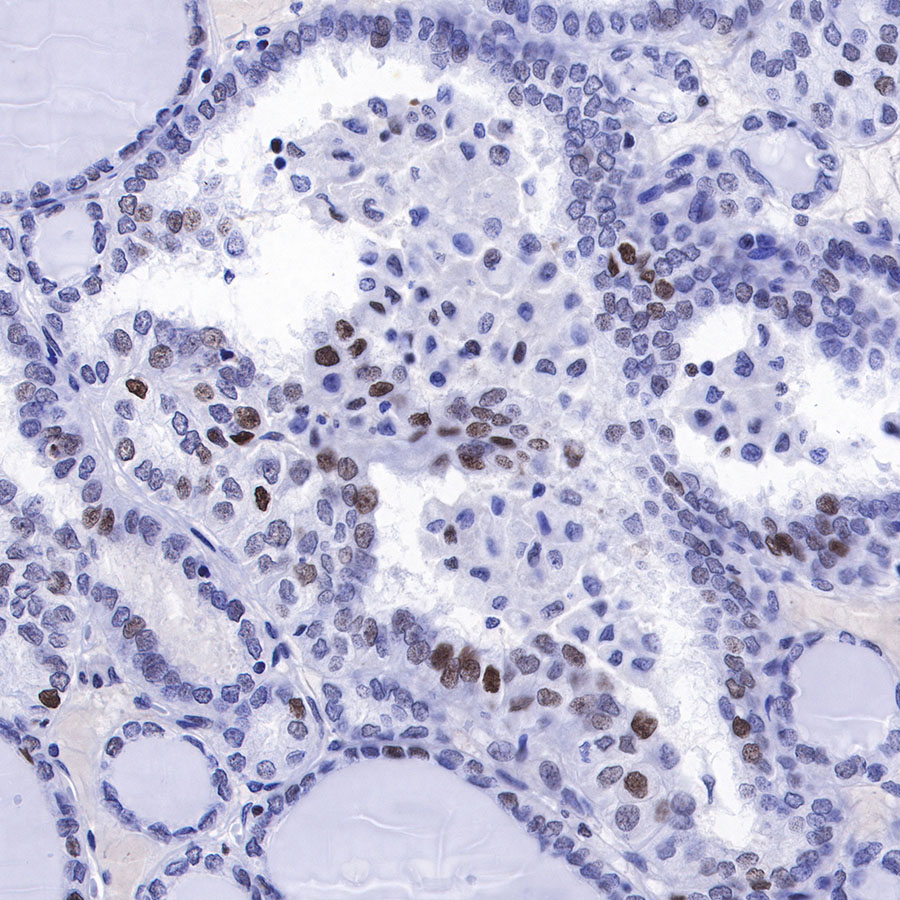
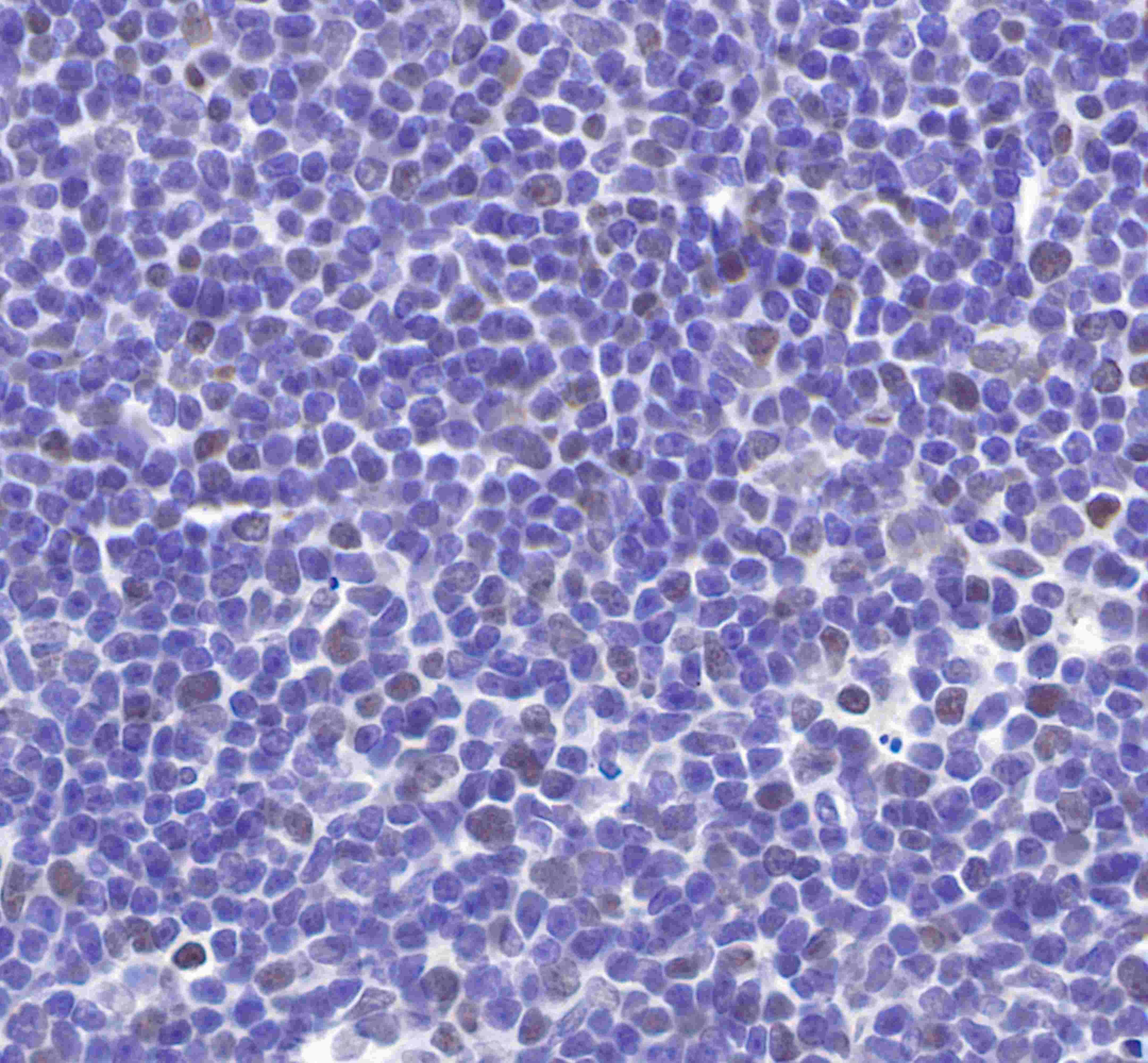
| IHC-P |
1:1000 |
|
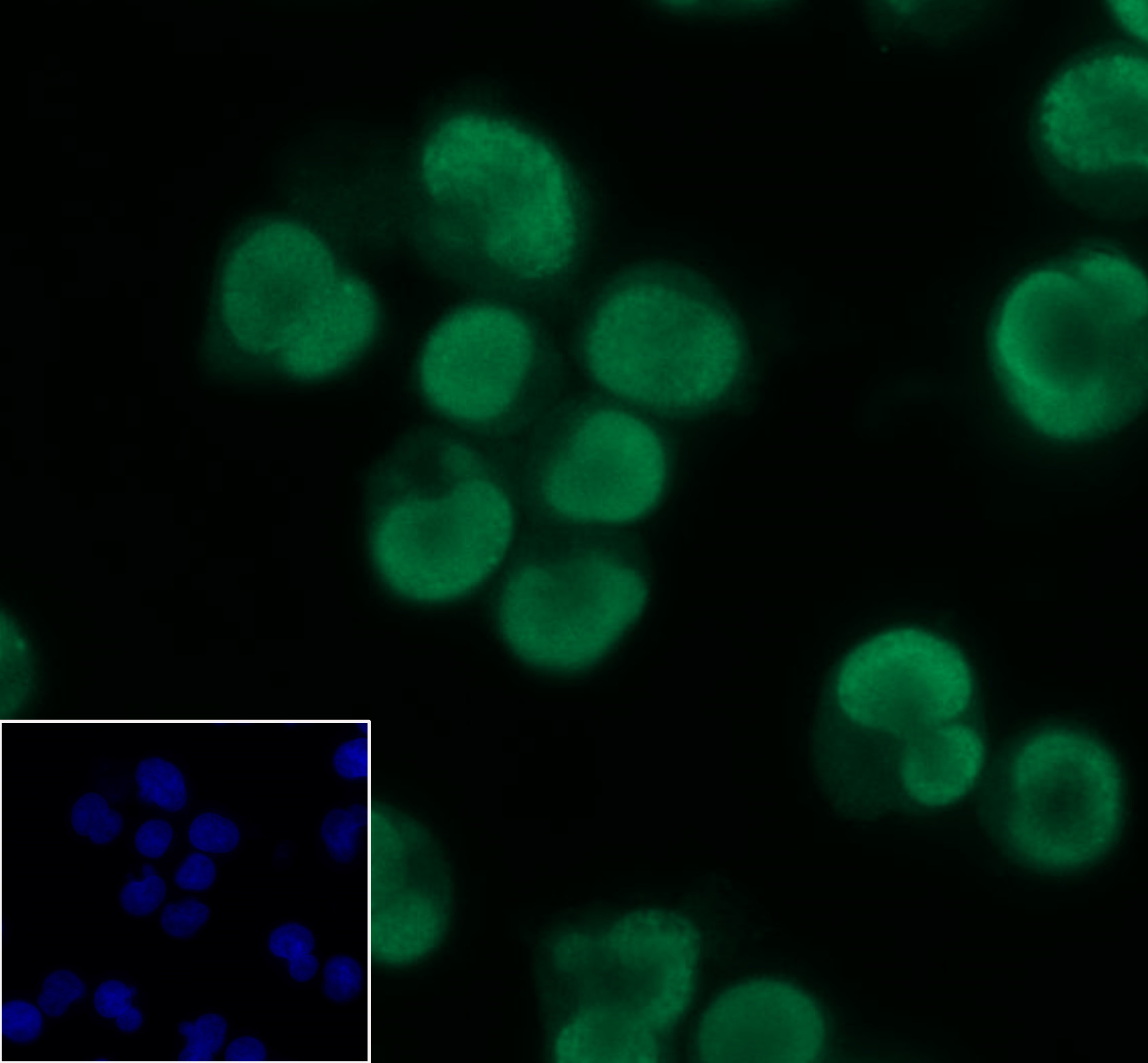
| ICC |
1:100 |
|
| WB |
1:1000 |
|
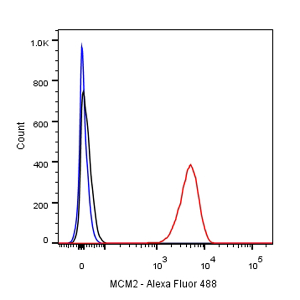
| ICFCM |
1:500 |
|
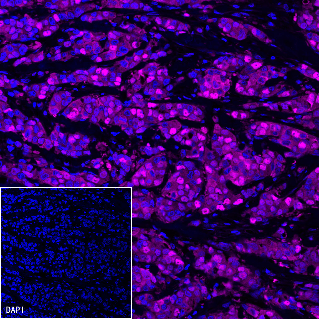
| IF |
1:500 |
|
Background
MCM2 is one of the highly conserved mini-chromosome maintenance proteins (MCM) that are involved in the initiation of eukaryotic genome replication. The hexameric protein complex formed by MCM proteins is a key component of the pre-replication complex (pre-RC) and may be involved in the formation of replication forks and in the recruitment of other DNA replication related proteins. MCM2 forms a complex with MCM4, 6, and 7, and has been shown to regulate the helicase activity of the complex. MCM2 is phosphorylated, and thus regulated by protein kinases CDC2 and CDC7.