Product Specification
| Host |
Rabbit |
| Antigen |
E-Cadherin |
| Synonyms |
Cadherin-1, CD324 |
| Immunogen |
N/A |
| Location |
Cell membrane |
| Accession |
P12830 |
| Clone Number |
SDT-R088 |
| Antibody Type |
Rabbit mAb |
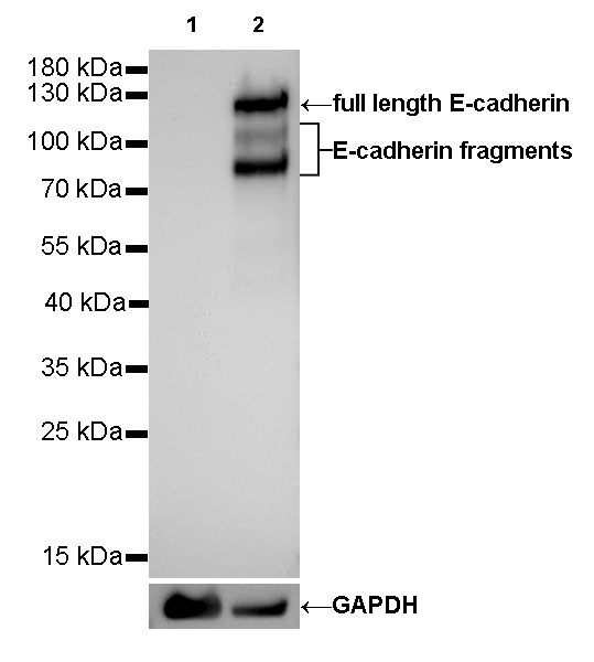
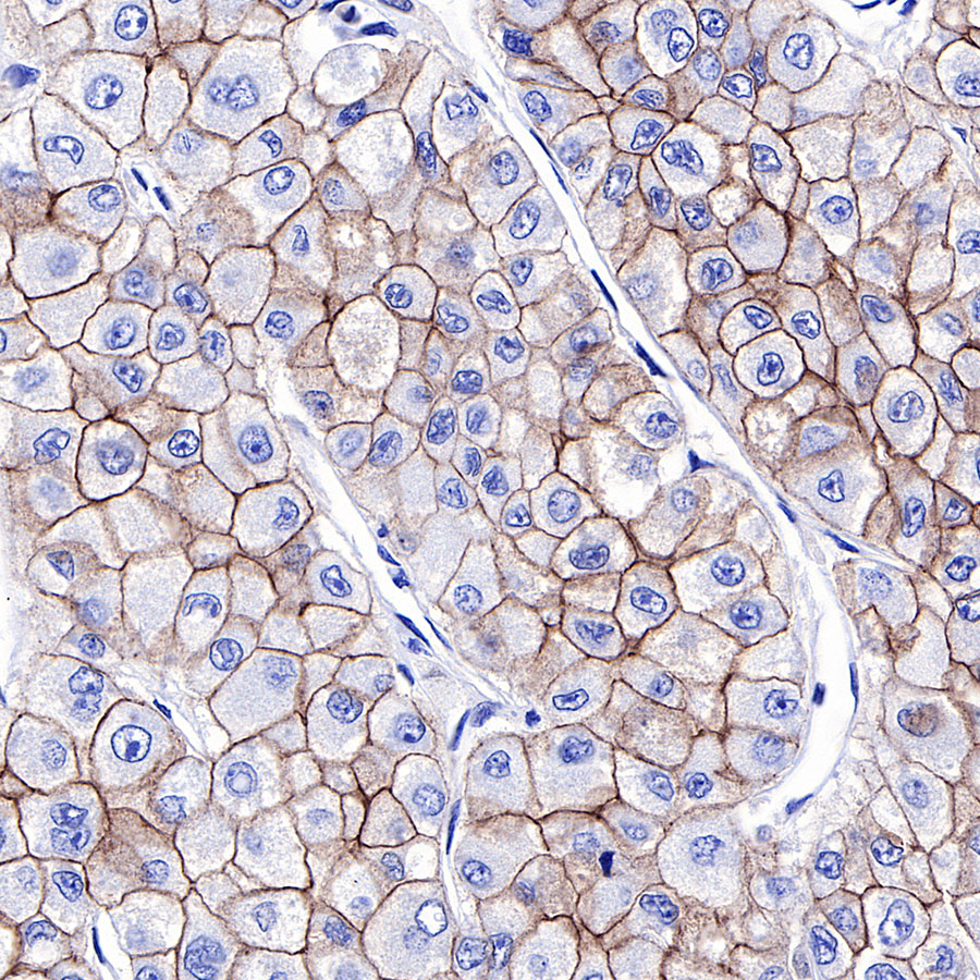
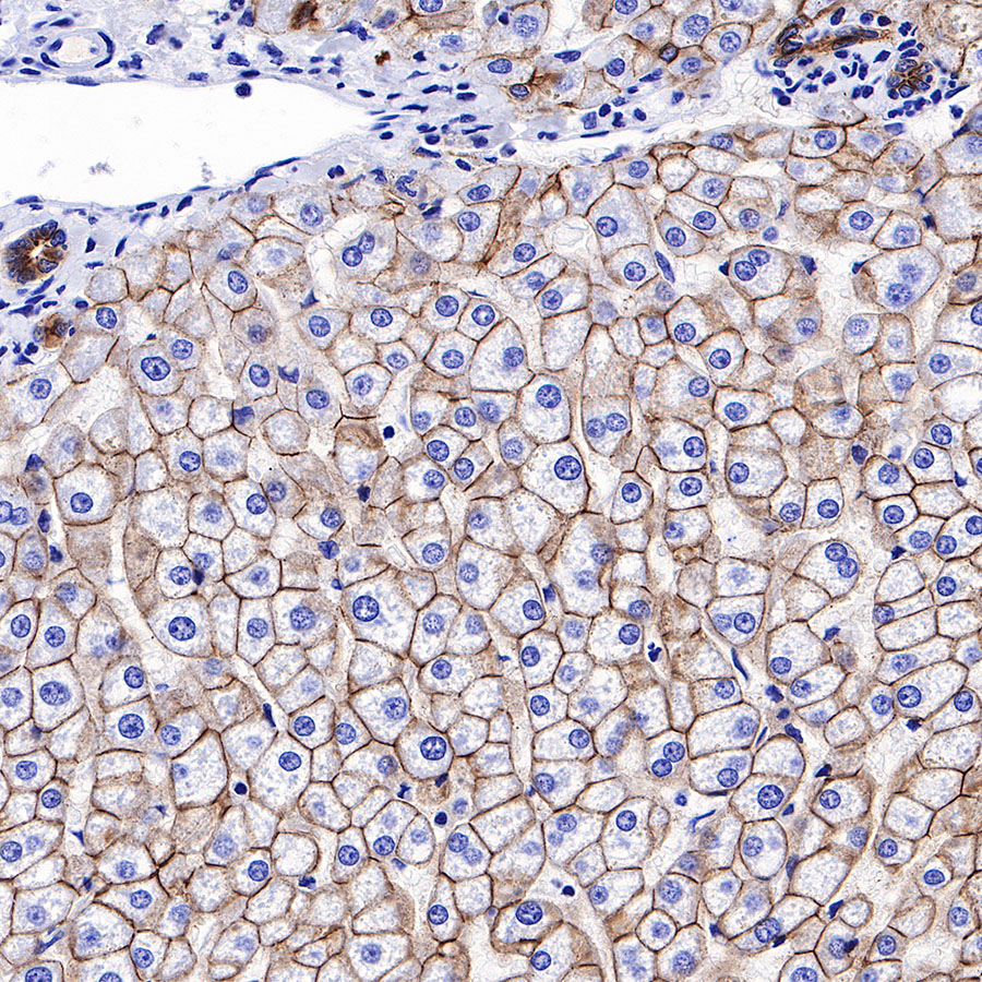
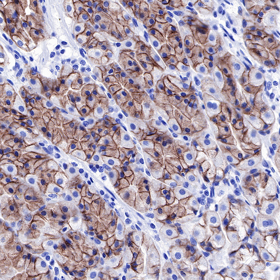
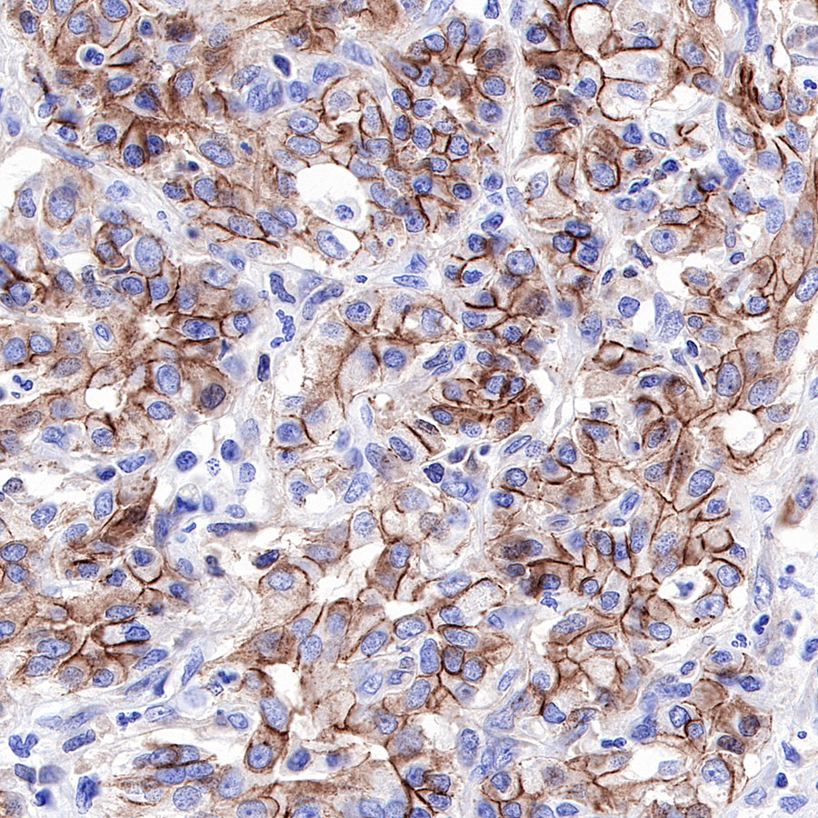
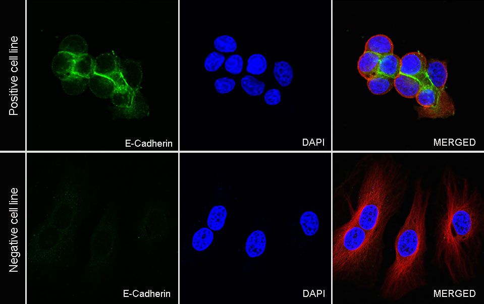
| Application |
WB, IHC-P, ICC |
| Reactivity |
Hu |
| Purification |
Protein A |
| Concentration |
0.25 mg/ml |
| Physical Appearance |
Liquid |
| Storage Buffer |
PBS, 40% Glycerol, 0.05%BSA, 0.03% Proclin 300 |
| Stability & Storage |
12 months from date of receipt / reconstitution, -20 °C as supplied |
Dilution
| application |
dilution |
species |
| IHC-P |
1:1000 |
|
| WB |
1:500 |
|
| ICC |
1:100 |
|
Background
E-cadherin is a transmembrane glycoprotein which connects epithelial cells together at adherens junctions. In normal cells, E-cadherin exerts its tumour suppressing role mainly by sequestering β-catenin from its binding to LEF (Lymphoid enhancer factor)/TCF (T cell factor) which serves the function of transcribing genes of the proliferative Wnt signaling pathway. Despite the ongoing debate on whether the loss of E-cadherin is the cause or effect of epithelial-mesenchymal transition (EMT), E-cadherin functional loss has frequently been associated with poor prognosis and survival in patients of various cancers. The dysregulation of E-cadherin expression that leads to carcinogenesis happens mostly at the epigenetic level but there are cases of genetic alterations as well. E-cadherin expression has been linked to the cellular functions of invasiveness reduction, growth inhibition, apoptosis, cell cycle arrest and differentiation.