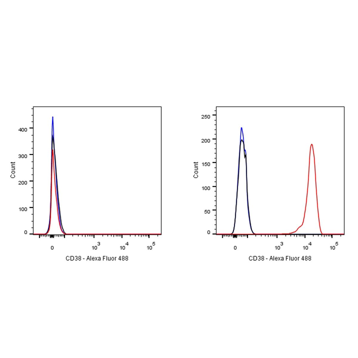
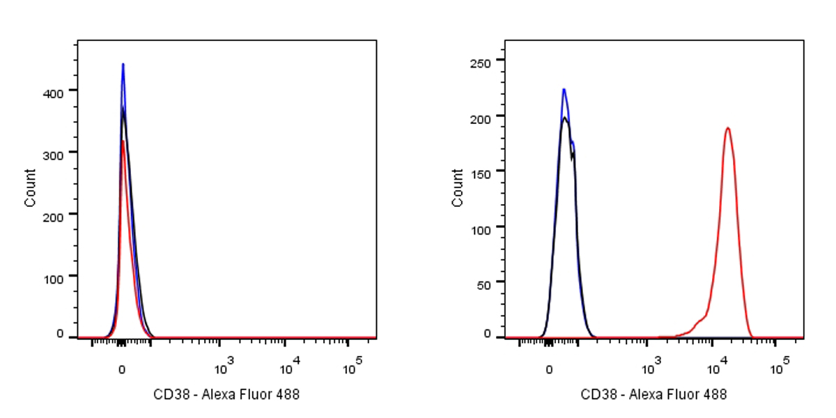
Flow cytometric analysis of HeLa (left) / Ramos (right) cells labelling CD38 antibody at 1/50 dilution (0.1ug)/ (red) compared with a Rabbit monoclonal IgG (Black) isotype control and an unlabelled control (cells without incubation with primary antibody and secondary antibody) (Blue). Goat Anti-Rabbit IgG Alexa Fluor® 488 at 1/1000 dilution was used as the secondary antibody. Negative control: HeLa
S-RMab® CD38 Recombinant Rabbit mAb (SDT-031-45-2)
S-RMab® CD38 Recombinant Rabbit mAb (SDT-031-45-2)
Price:
Regular price
$100 USD
Regular price
Sale price
$100 USD
Unit price
per
For shipping services or bulk orders, you may request a quotation.
Secure checkout with

View full details
Product Details
Product Details
Product Specification
| Host | Rabbit |
| Antigen | CD38 |
| Synonyms | 2'-phospho-ADP-ribosyl cyclase, 2'-phospho-ADP-ribosyl cyclase/2'-phospho-cyclic-ADP-ribose transferase,2'-phospho-cyclic-ADP-ribose transferase,ADP-ribosyl cyclase 1 (ADPRC 1),Cyclic ADP-ribose hydrolase 1 (cADPr hydrolase 1),T10 |
| Immunogen | Synthetic Peptide |
| Location | Membrane |
| Accession | P28907 |
| Clone Number | SDT-031-45-2 |
| Antibody Type | Rabbit mAb |
| Application | WB, IHC-P, ICC, FC |
| Reactivity | Hu |
| Purification | Protein A |
| Concentration | 0.05mg/ml |
| Conjugation | Unconjugated |
| Physical Appearance | Liquid |
| Storage Buffer | PBS, 40% Glycerol, 0.05%BSA, 0.03% Proclin 300 |
| Stability & Storage | 12 months from date of receipt / reconstitution, -20 °C as supplied |
Dilution
| application | dilution | species |
| FC | 1:50 | null |
| IHC-P | 1: 500-1:1000 | null |
| WB | 1:500 | null |
| ICC | 1:50 | null |
Background
CD38 (cluster of differentiation 38), also known as cyclic ADP ribose hydrolase is a glycoprotein found on the surface of many immune cells (white blood cells), including CD4+, CD8+, B lymphocytes and natural killer cells. CD38 also functions in cell adhesion, signal transduction and calcium signaling. The loss of CD38 function is associated with impaired immune responses, metabolic disturbances, and behavioral modifications including social amnesia possibly related to autism. The CD38 protein is a marker of cell activation. It has been connected to HIV infection, leukemias, myelomas, solid tumors, type II diabetes mellitus and bone metabolism, as well as some genetically determined conditions.
Picture
Picture
Validation Data
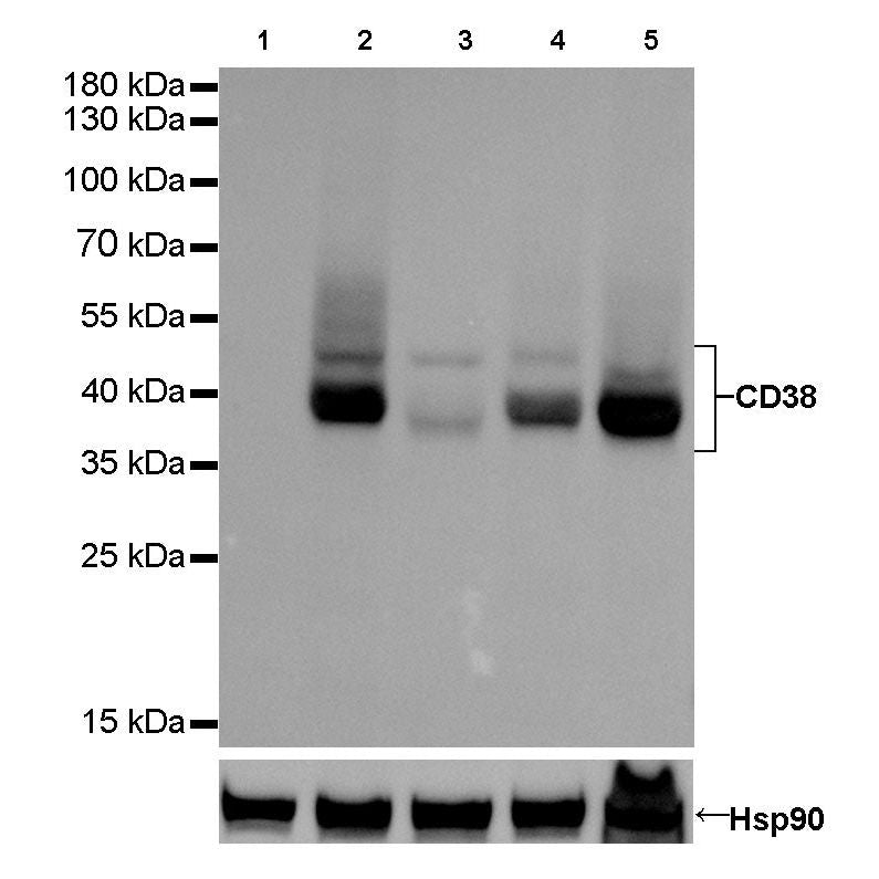
Western Blot
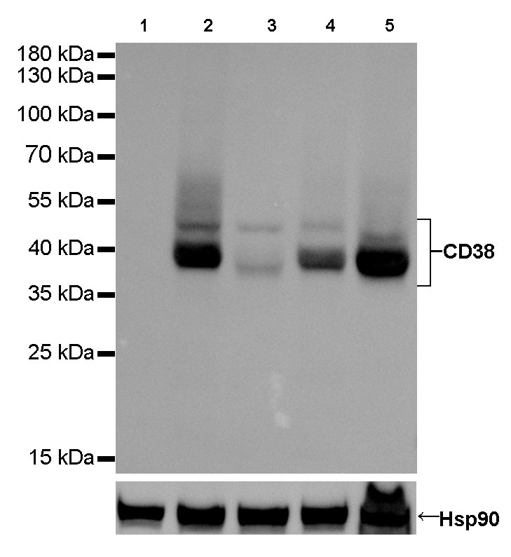
WB result of CD38 Rabbit mAb
Primary antibody : CD38 Rabbit mAb at 1/500 dilution
Lane 1: HepG2 whole cell lysate 20 µg
Lane 2: Daudi whole cell stomach lysate 20 µg
Lane 3: Jurkat whole cell lysate 20 µg
Lane 4: Raji whole cell heart lysate 20 µg
Lane 5: Ramos whole cell heart lysate 20 µg
Negative control: HepG2 whole cell lysate
Secondary antibody: Goat Anti-Rabbit IgG, (H+L), HRP conjugated at 1/10000 dilution
Predicted MW: 34 kDa
Observed MW: 40kDa
Exposure time: 1.3 s
Immunohistochemistry
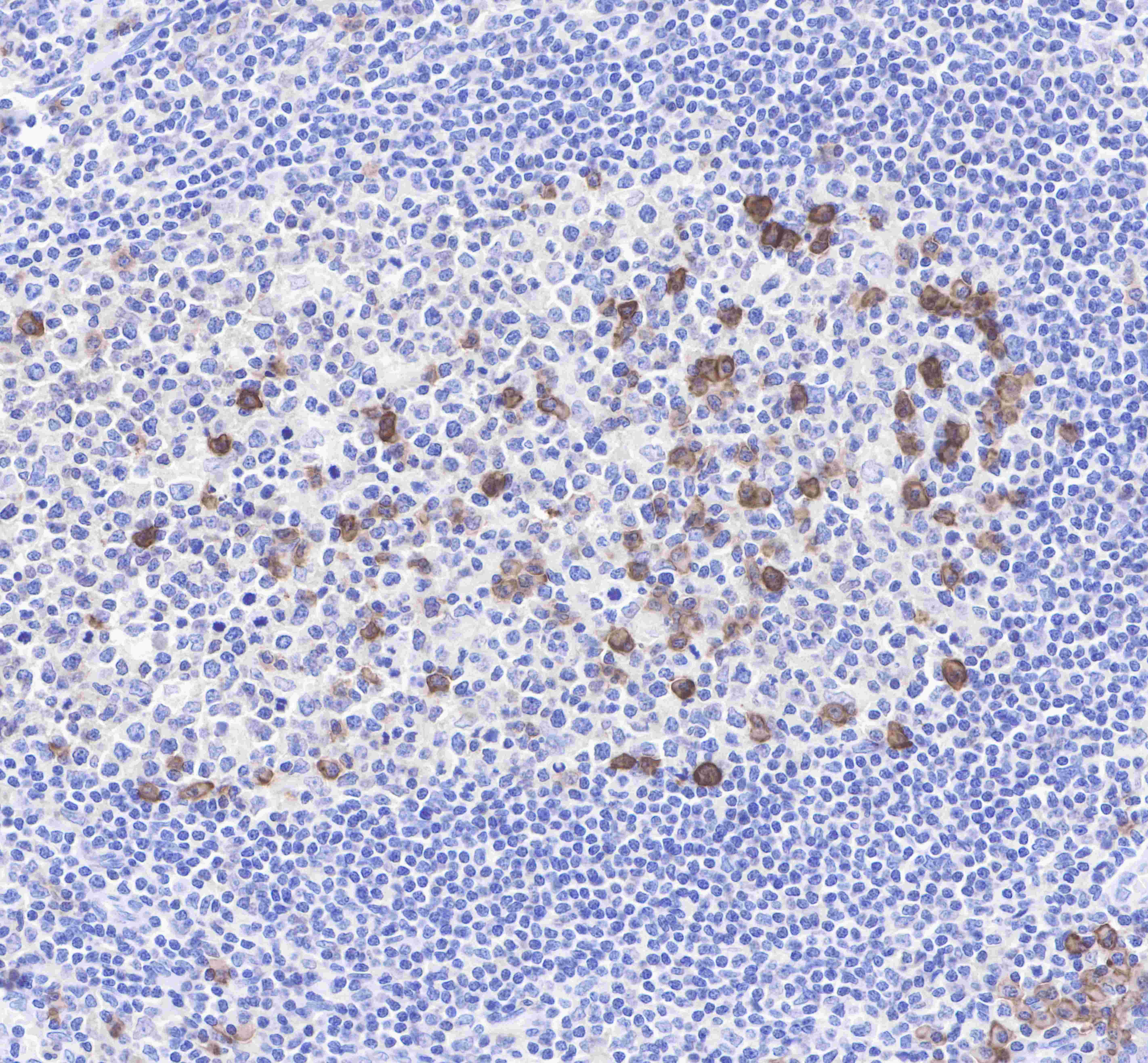
IHC shows positive staining in paraffin-embedded human tonsil. Anti-CD38 antibody was used at 1/1000 dilution, followed by a Goat Anti-Rabbit IgG H&L (HRP) ready to use. Counterstained with hematoxylin.
Heat mediated antigen retrieval with Tris/EDTA buffer pH9.0 was performed before commencing with IHC staining protocol.
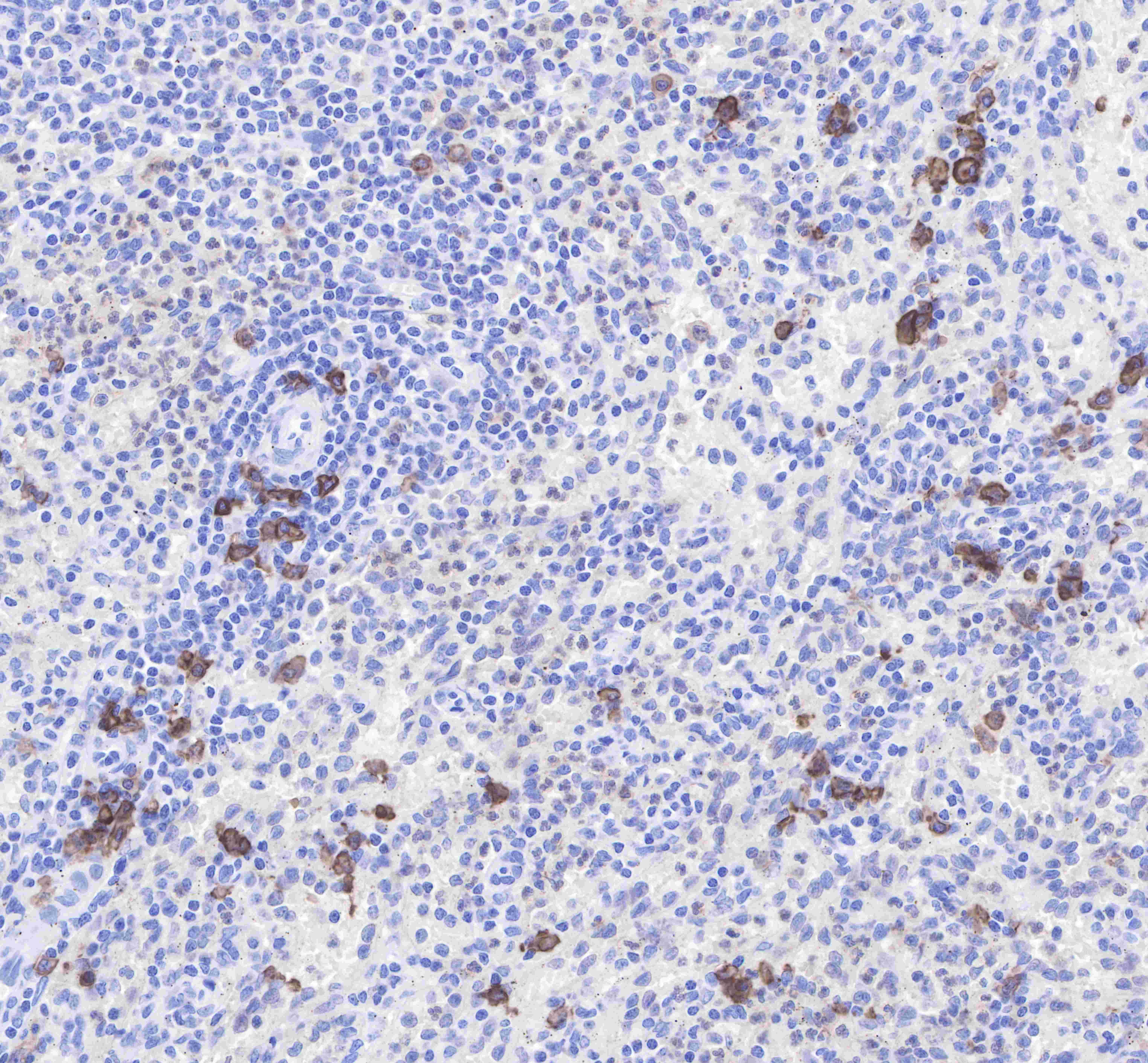
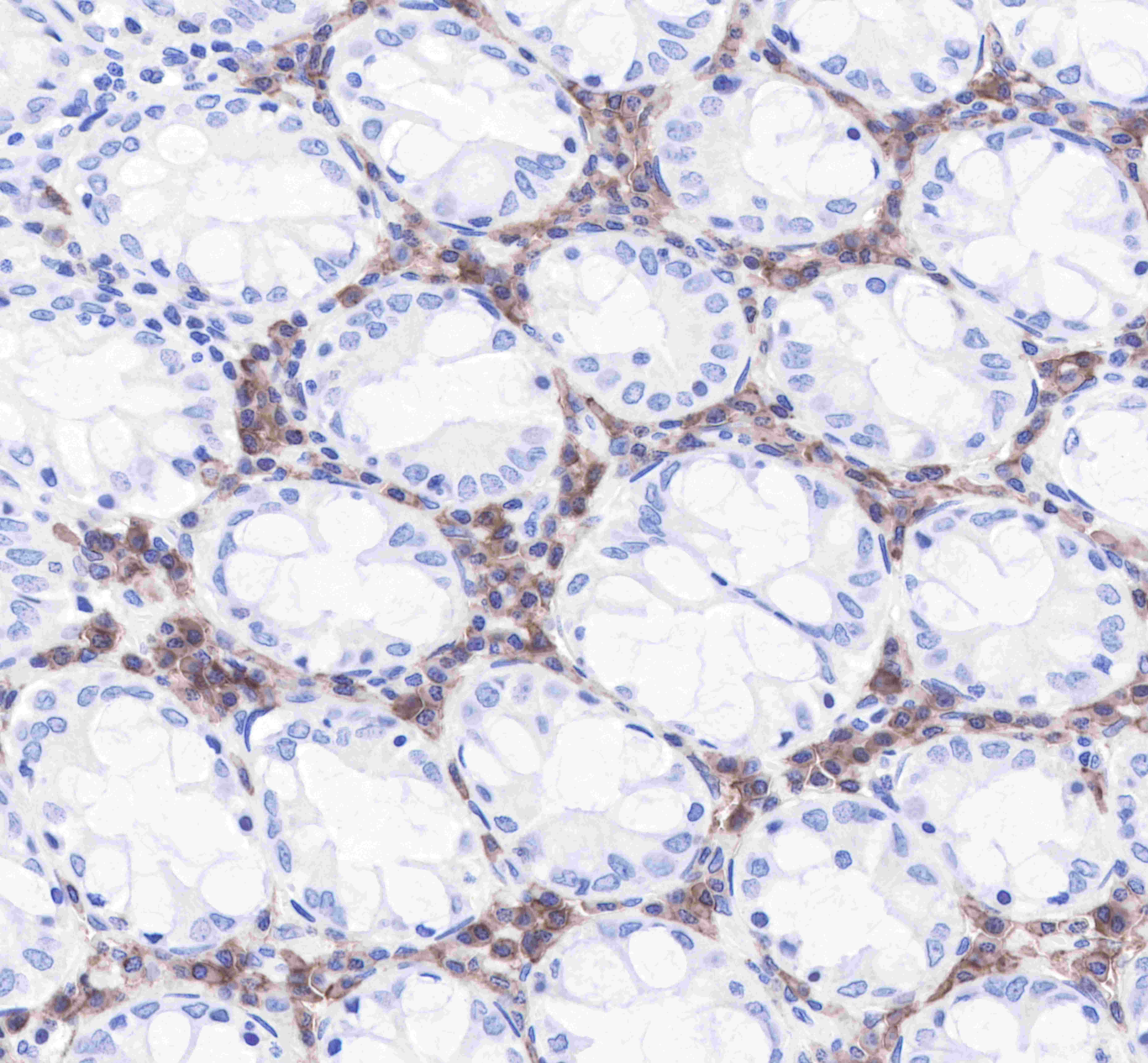
IHC shows positive staining in paraffin-embedded human spleen. Anti-CD38 antibody was used at 1/1000 dilution, followed by a Goat Anti-Rabbit IgG H&L (HRP) ready to use. Counterstained with hematoxylin.
Heat mediated antigen retrieval with Tris/EDTA buffer pH9.0 was performed before commencing with IHC staining protocol.
IHC shows positive staining in paraffin-embedded human colon. Anti-CD38 antibody was used at 1/1000 dilution, followed by a Goat Anti-Rabbit IgG H&L (HRP) ready to use. Counterstained with hematoxylin.
Heat mediated antigen retrieval with Tris/EDTA buffer pH9.0 was performed before commencing with IHC staining protocol.
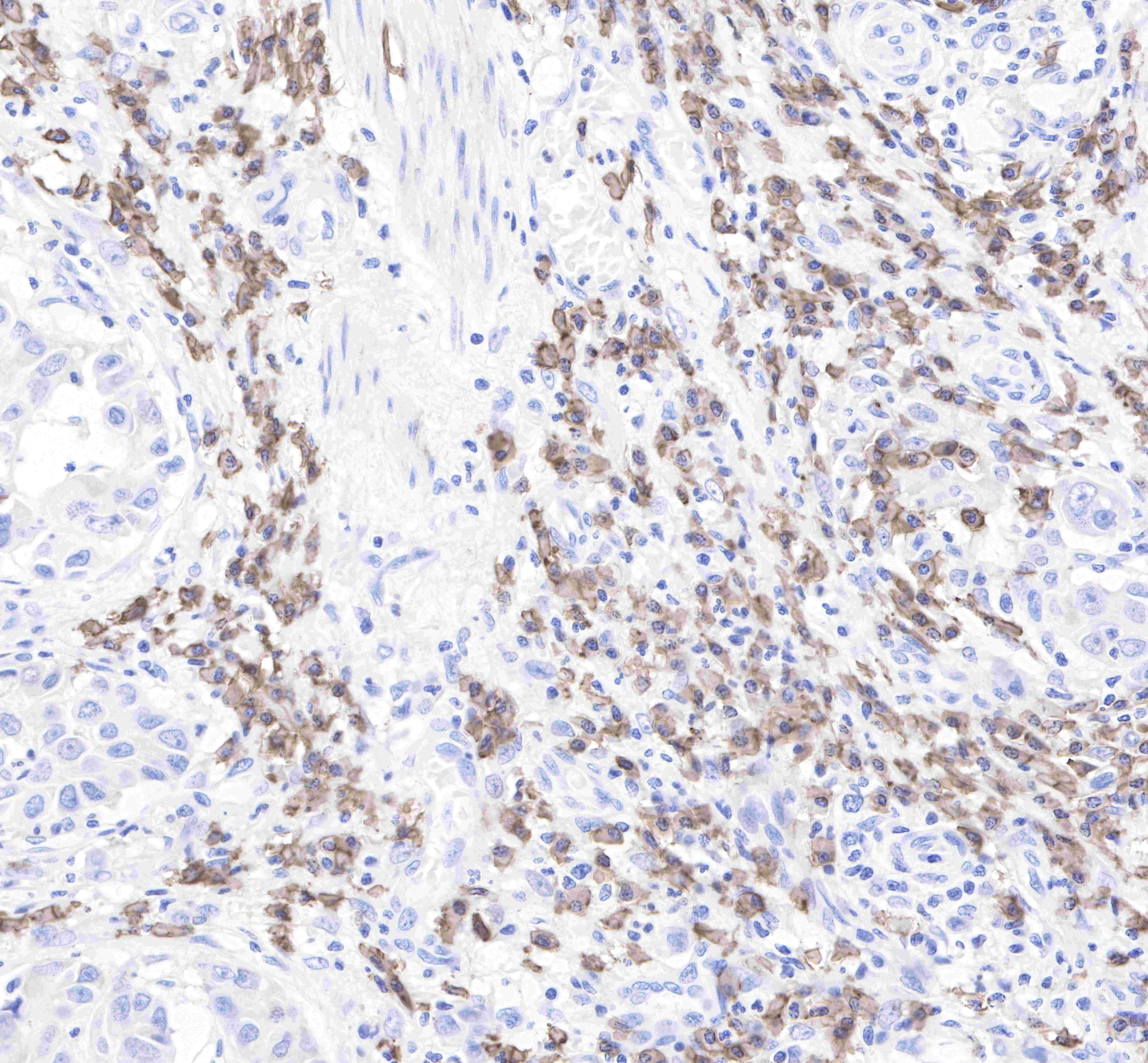
IHC shows positive staining in paraffin-embedded human stomach. Anti-CD38 antibody was used at 1/1000 dilution, followed by a Goat Anti-Rabbit IgG H&L (HRP) ready to use. Counterstained with hematoxylin.
Heat mediated antigen retrieval with Tris/EDTA buffer pH9.0 was performed before commencing with IHC staining protocol.
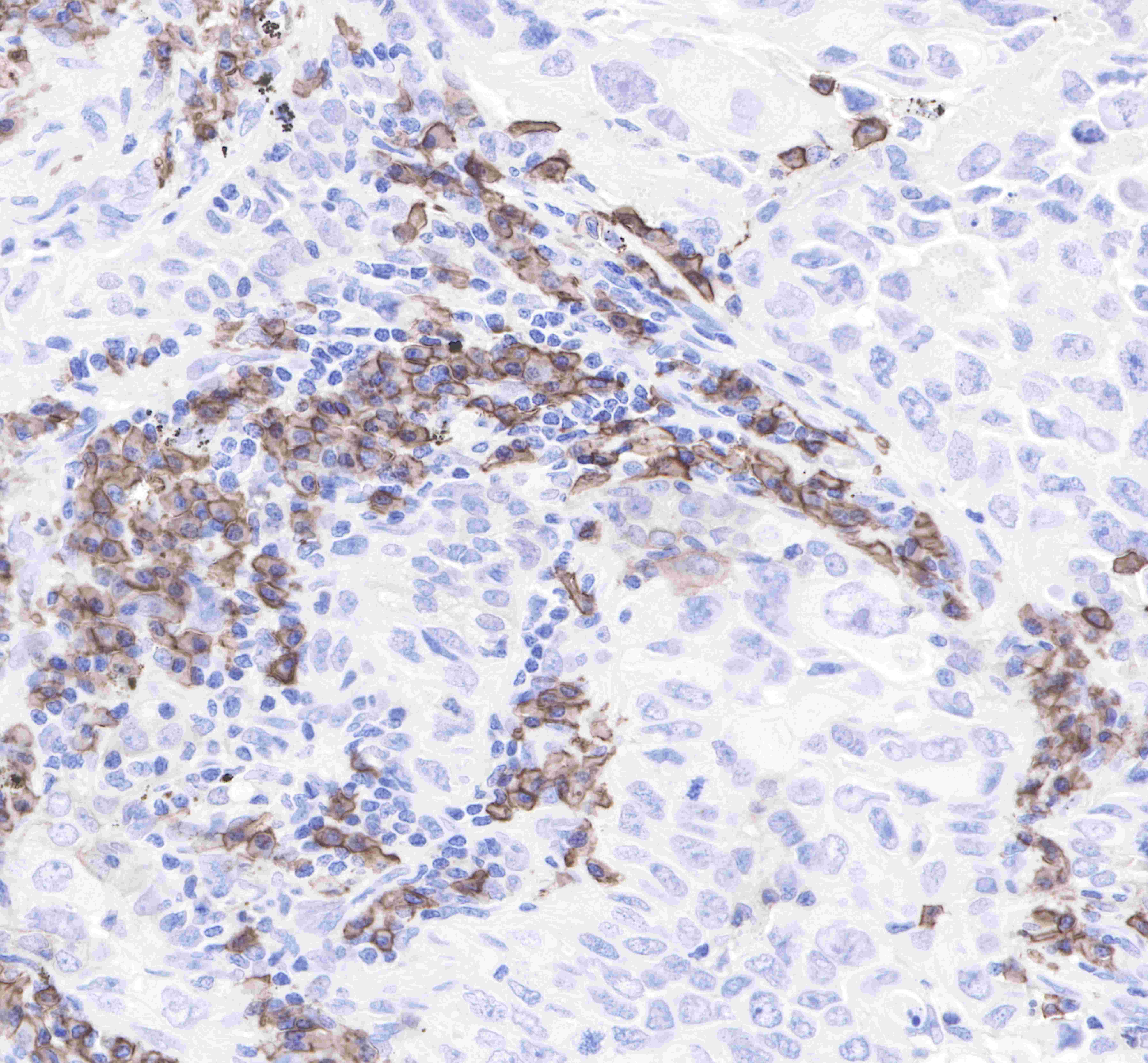
IHC shows positive staining in paraffin-embedded human lung adenocarcinoma. Anti-CD38 antibody was used at 1/1000 dilution, followed by a Goat Anti-Rabbit IgG H&L (HRP) ready to use. Counterstained with hematoxylin.
Heat mediated antigen retrieval with Tris/EDTA buffer pH9.0 was performed before commencing with IHC staining protocol.
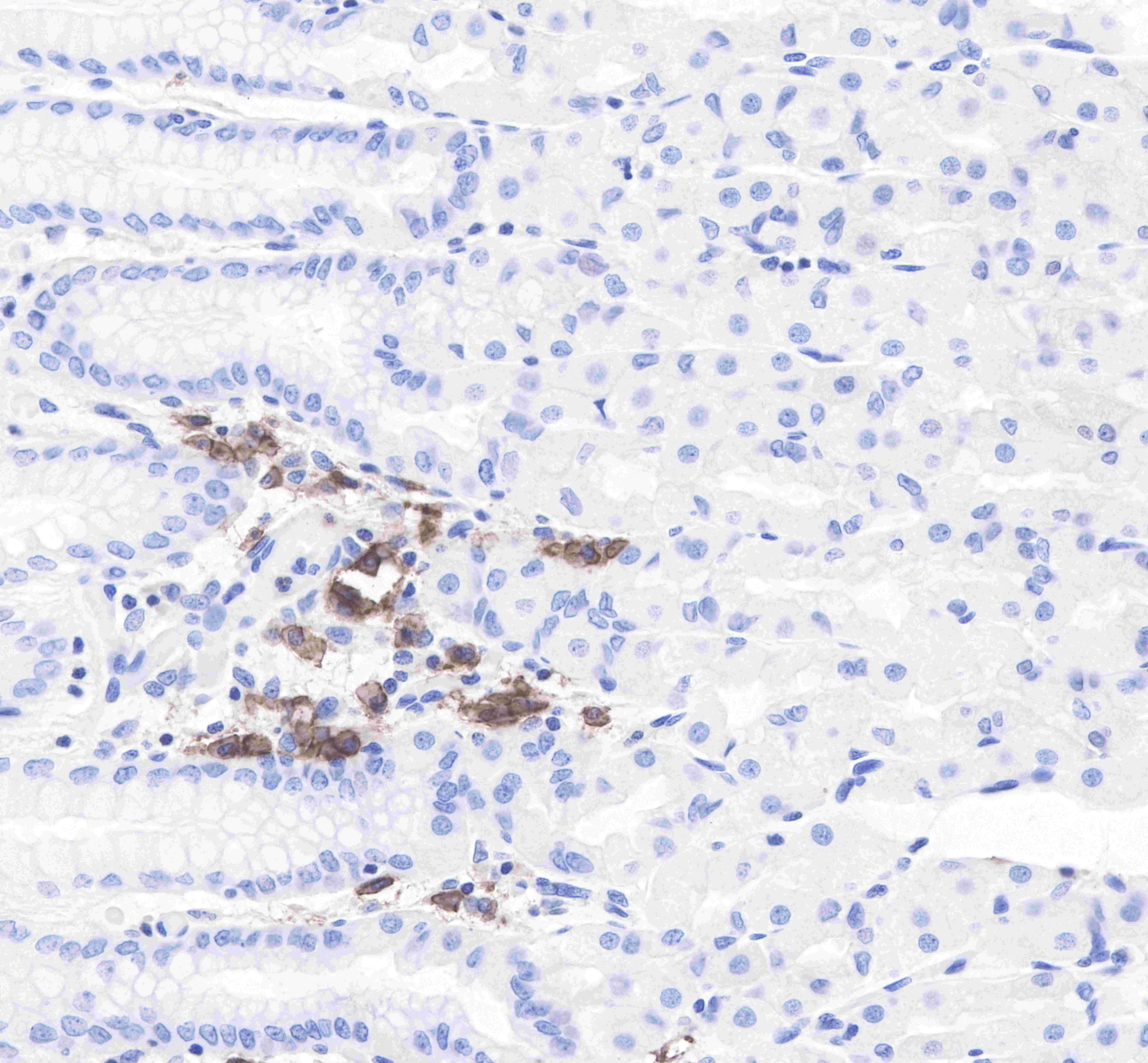
IHC shows positive staining in paraffin-embedded human prostate cancer. Anti-CD38 antibody was used at 1/1000 dilution, followed by a Goat Anti-Rabbit IgG H&L (HRP) ready to use. Counterstained with hematoxylin.
Heat mediated antigen retrieval with Tris/EDTA buffer pH9.0 was performed before commencing with IHC staining protocol.
Immunocytochemistry

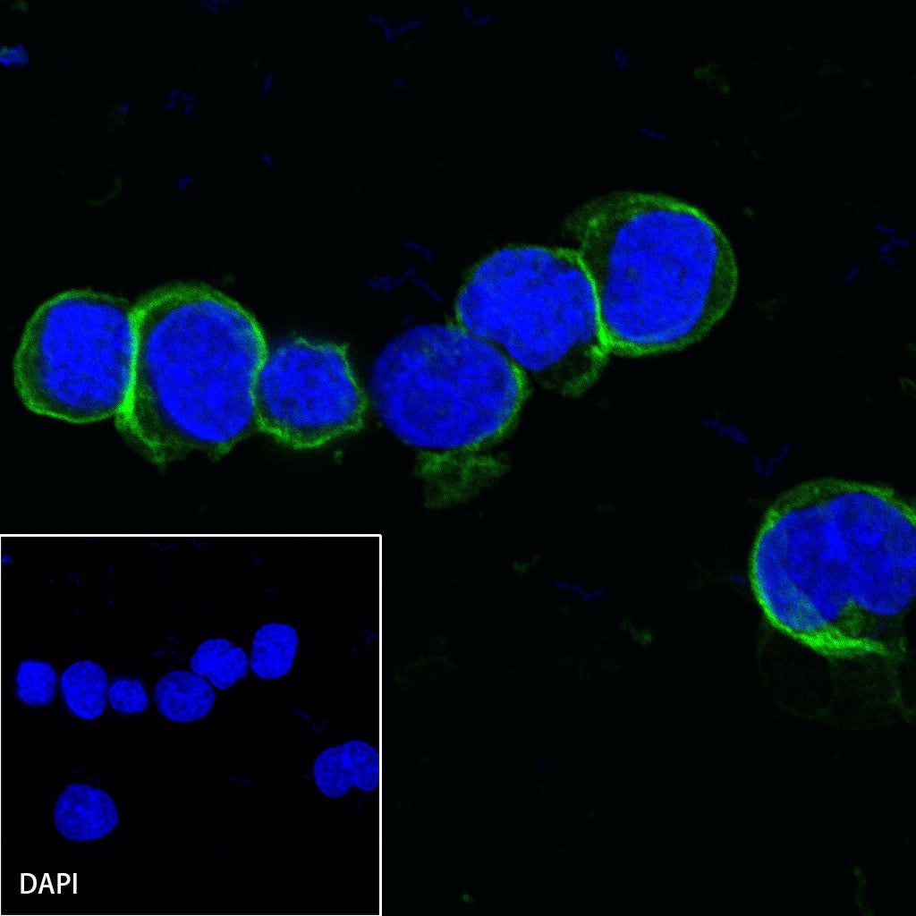
ICC shows positive staining in Ramos cells. Anti-CD38 antibody was used at 1/50 dilution (Green) and incubated overnight at 4°C. Goat polyclonal Antibody to Rabbit IgG - H&L (Alexa Fluor® 488) was used as secondary antibody at 1/1000 dilution. The cells were fixed with 4% PFA and permeabilized with 0.1% PBS-Triton X-100. Nuclei were counterstained with DAPI (Blue).
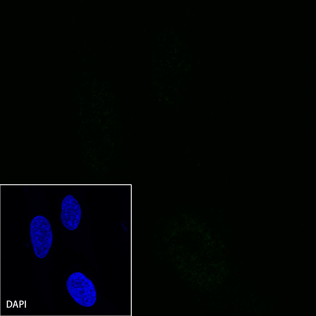
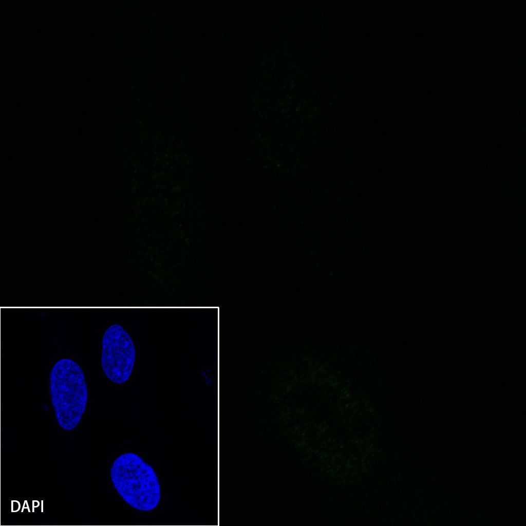
Negative control:ICC shows negative staining in HeLa cells. Anti-CD38 antibody was used at 1/50 dilution and incubated overnight at 4°C. Goat polyclonal Antibody to Rabbit IgG - H&L (Alexa Fluor® 488) was used as secondary antibody at 1/1000 dilution. The cells were fixed with 4% PFA and permeabilized with 0.1% PBS-Triton X-100. Nuclei were counterstained with DAPI (Blue).