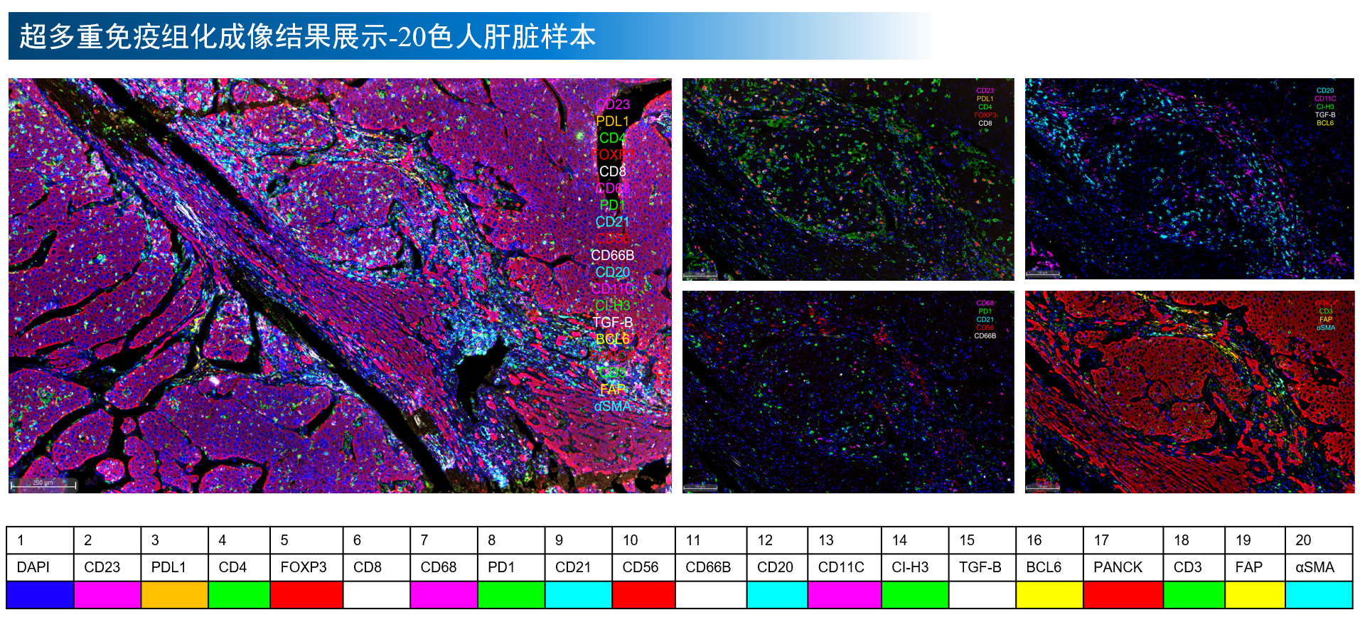
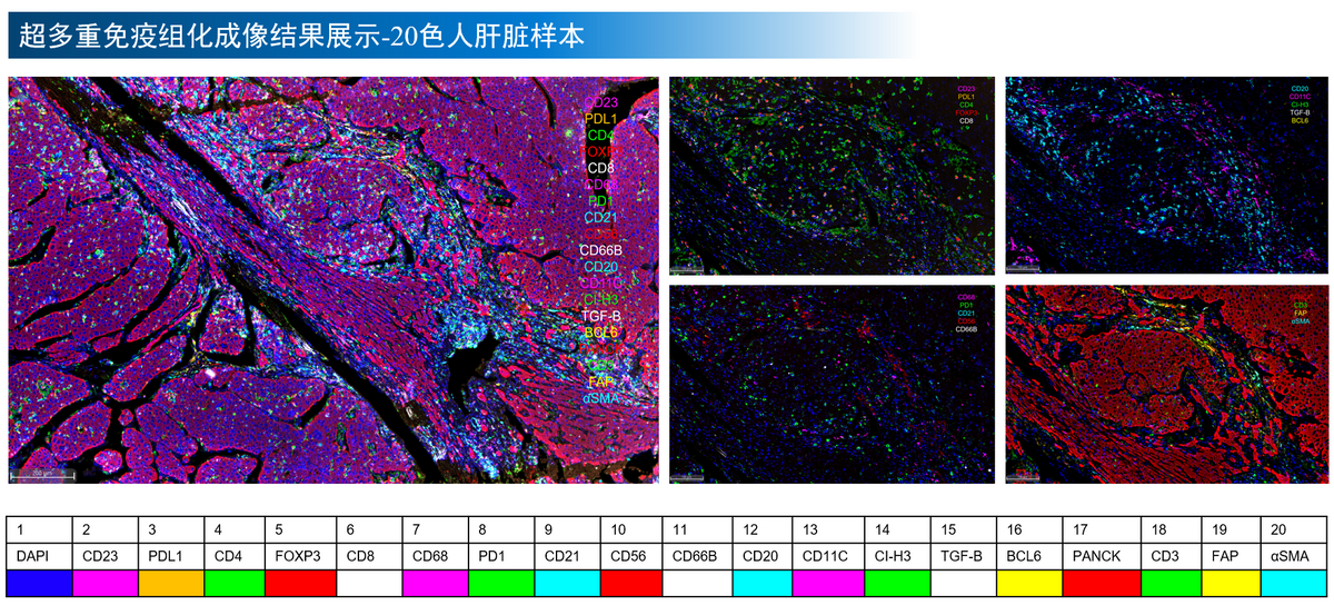
19-labeled 20-color supermultiplex immunohistochemical imaging results of human liver section samples
HyperTSA Ultra Multiplex IHC Kit
HyperTSA Ultra Multiplex IHC Kit
Price:
Regular price
$1,246 USD
Regular price
Sale price
$1,246 USD
Unit price
per
For shipping services or bulk orders, you may request a quotation.
Secure checkout with

View full details
Product Details
Product Details
Product Specification
| Usage |
1. Detection sample 1 biopsy or surgically removed samples of different species; 2 Use after tissue isolation 10% Neutral formalin fixed, fixed time 8-48 h Best; 3 Recommended tissue section thickness 3-4 μm Use an anti-fall slide. 2. Operation mode Hand dyeing 3. Required experimental equipment, consumables and reagents 1 Experimental equipment: Incubator, microwave oven, centrifuge, vortex mixer 2 Experimental consumables: Dyeing cylinder, incubation wet box, drying plate, anti-slide slide, cover slide, washing bottle, measuring cylinder, pipette, immunohistochemistry hydrophobic pen, timer, EP Tube, pipette tip, blue cap vial 3 Experimental reagents: Sterilized deionized water, xylene, ethanol ( 100% 、 95% 、 75% )、 10% Neutral formalin, anti-fluorescence quenching sealant, Tween -20 , Triton 4. Working fluid preparation 1 Fluorescent dye working solution The dye in the kit is 100X Mother liquor, placed at room temperature before use 10min , reuse HyperTSA Signal amplification solution according to 1:100 Dilute and use (configure within half an hour before use, do not configure in advance). 2 、 HyperTSA HRP Enzyme-labeled anti-mouse / Rabbit IgG Polymer secondary antibody working solution (3x) With buffer ( TBS 、 PBS 、 TBST 、 PBST Either can) will 3X Enzyme-labeled anti-mouse / Rabbit IgG The polymer secondary antibody concentrate is diluted and prepared as a secondary antibody working solution (prepared within half an hour before use, do not prepare in advance). 3 、 HyperTSA DAPI Working fluid As per drop DAPI Concentrate plus 500 μL Ratio of buffer, dropwise HyperTSA DAPI Concentrate solution into buffer solution, mix well to obtain DAPI Working fluid. 4 Antigen recovery solution working solution Use deionized water to 50X The concentrate was prepared according to 1:50 The dilution was configured as an antigen retrieval working solution. 5 、 HyperTSA Fluorescent Eluent Working Solution ( 5X ) Use deionized water to 5X The concentrate dilution was configured as a fluorescent eluent working solution. 6 、 TBST In TBS Add in 0.1% Twain of -20 And 0.03% of Triton Configured as working fluid. V. Experimental operation Note: Before starting the experiment, please confirm that other equipment, consumables, reagents, etc. required for the experiment have been prepared. Carefully read the [Working Solution Preparation] section and prepare the working solution as required. 1 The paraffin sections are placed in 60℃ Baking in incubator 60 min ; 2 , dewaxing and hydration: xylene (10 min)→ Xylene (10 min) → Xylene (10 min)→ Anhydrous ethanol (5 min×2 Secondary )→95% Ethanol (5 min×1)→75% Ethanol (5 min) (You can also choose environmentally friendly tissue transparent liquid, refer to its instructions for specific operation) ; 3 Distilled water flushing 5 min×2 Secondary; 4 Antigen remediation: immerse the slice in the antigen remediation solution working liquid for microwave remediation and preheat 5 min , high fire 2 min , low fire 15 min ; 5 Natural cooling at room temperature; 6 Washing: Use TBST washing 3 Secondary , 5 min/ Secondary; First round of staining 7 Blocking: Blocking solution is blocked at room temperature 15 min ; 8 Primary antibody incubation: dropwise addition of primary antibody working solution 100 μl , 37℃ incubation 1 h (or 4℃ overnight incubation); 9 Washing: TBST washing 3 times, 5 min/ Secondary; 10 Secondary antibody incubation: dropwise addition of secondary antibody 100 μl , 37℃ incubation 10 min ; 11 Washing: TBST washing 3 Secondary , 5 min/ Secondary; 12 Fluorescence color development: dropwise addition of fluorescent dye working solution 100 μl , room temperature 5 min; 13 Washing: TBST washing 3 times, 5 min/ Secondary; 14 Microwave treatment: repeating the steps 4-6 ; 15 Sequential antibody staining: the first antibody staining is completed, and each subsequent antibody needs to repeat the steps 7-14 Complete all antibody staining sequentially; 16 dropwise addition DAPI Working fluid 100 μl Staining, room temperature 5 min ; 17 Washing: distilled water washing 3 times, 5 min/ Secondary; 18 Dropwise adding antifluorescence quenching sealing agent 100 μl , coverslip seal scan. 19 After the first round of staining image scanning is completed, the slide is soaked in buffer solution to remove the coverslip (the soaking time varies according to the length of the scanning time of the seal). 20 Elution step: preheating the fluorescence eluent working liquid in a microwave oven over high heat 3 min Immersing the sections in the fluorescent eluent working fluid, microwave repair, low fire 15 min ; 21 Natural cooling at room temperature; 22 Washing: Use TBST washing 3 Secondary , 5 min/ Secondary; Second round of staining 23 Repeat 7-18 Step 2 completes the second round of image acquisition. If necessary, multiple rounds of subsequent staining should be completed in sequence. 24 Perform single-round image analysis as needed or register multiple rounds of images using software for super-multi-target analysis. * All the conditions in the experimental procedure are recommended, and the specific experimental conditions are adjusted according to the actual situation. |
||||||||||||||||||||||||||||||||||||||||||||||||||||||||||||||||||
| Synonym | Hypermultiplex immunohistochemical staining kit | ||||||||||||||||||||||||||||||||||||||||||||||||||||||||||||||||||
| Description | HyperTSA washable ultra-multiplex immunohistochemical staining kit is a scientific research tool for tissue space in situ detection. It innovatively combines tyramine signal amplification (TSA) technology with elutable technology. After each round of multiplex staining and imaging, the covalently bound fluorescent dye is eluted by elutable technology for the next round of multiplex staining and imaging. Image registration of multiple rounds of staining data can realize in situ labeling of supermultiplex protein targets in one slice, breaking through the limitation of traditional TSA technology (7-9 targets), and realizing the detection of 10-50 protein markers. Through the cyclic workflow of "staining-imaging-elution-staining", it can not only save tissue samples, but also reveal the spatial interaction relationship of cell subsets in complex tissues, which provides strong technical support for cutting-edge fields such as tumor microenvironment research and neural circuit analysis. Principle of dyeing: HyperTSA ultra-multiplex immunohistochemical staining kit innovatively integrates tyramine signal amplification (TSA) technology and reversible staining technology, providing researchers with an efficient and flexible multi-target detection solution. The core of the kit contains four kinds of HyperTSA fluorescent dyes with excellent spectral characteristics. With special signal amplification solution and high-efficiency polymer secondary antibody, it can realize accurate co-localization analysis of more than 10 protein markers. Its complete working system also includes: tissue antigen retrieval solution (suitable for multiple sample pretreatment needs), multifunctional blocking/primary antibody diluent (effective in reducing background interference), high-purity DAPI concentrate (nuclear localization labeling), and revolutionary fluorescent dye eluent (achieving gentle dye removal). In a single round of experiments, conventional TSA staining method can be used for multiplex staining. After the staining is completed, TSA fluorescent dye can be eluted with fluorescent dye eluent, so that the next round of staining can be carried out. After multiple rounds of staining, the detection of super-multiple targets on the same slice is realized. |
||||||||||||||||||||||||||||||||||||||||||||||||||||||||||||||||||
| Composition |
|
||||||||||||||||||||||||||||||||||||||||||||||||||||||||||||||||||
| General Notes | 1. PH value has an influence on dyeing. The slide should be clean and free of acid and alkali pollution, otherwise it will affect the dyeing effect. 2. Imaging the tissue in time after staining. If you need to store it, please avoid light. 3. Pay attention to gentle operation in the process of taking out the coverslip after each round of dyeing to reduce stripping. |
||||||||||||||||||||||||||||||||||||||||||||||||||||||||||||||||||
| Instructions | Hypermultiplex immunohistochemical staining for paraffin tissue sections | ||||||||||||||||||||||||||||||||||||||||||||||||||||||||||||||||||
| Storage Temp. | Long-term storage at-20℃, 2-8℃ after opening, sealed and protected from light. Valid for 12 months. |
Picture
Picture
Immunohistochemistry