Human IL-2 ELISA Kit
Human IL-2 ELISA Kit
Price:
Regular price
$450 USD
Regular price
Sale price
$450 USD
Unit price
per
For shipping services or bulk orders, you may request a quotation.
Secure checkout with

View full details
Product Details
Product Details
Product Specification
| Usage |
Need to bring your own test equipment 1. Microplate reader (measurable 450nm Absorption value of detection wavelength and 540nm Or 570nm Absorption value of corrected wavelength) 2. High precision liquid dispenser and disposable tip 3. Distilled or deionized water 4. Bottle washer (spray bottle), multi-channel plate washer or automatic plate washer 5. 500mL Measuring cylinder 1. Preparation before the experiment 1. Sample collection and storage ① Cell culture supernatant: particulate matter should be removed by centrifugation; Test the sample immediately. If the sample is not tested in time after collection, it is recommended to pack it according to the amount used once and store it frozen in -20℃ In the refrigerator, avoid repeated freezing and thawing. The sample may need to be used with a diluent ( 1× ) dilution. ② Serum: Use serum separation tubes ( SST ) Collect samples and place samples at room temperature 30 Minutes. Centrifugation 15 Minutes, with a rotation speed of 1000g 。 The serum was removed immediately and tested immediately. If the sample is not tested in time after collection, it is recommended to pack it according to the amount used once and store it frozen in ≤-20℃ In the refrigerator, avoid repeated freezing and thawing. The sample may need to be used with a diluent ( 1× ) dilution. ③ Plasma: Use EDTA , heparin or citric acid as an anticoagulant to collect plasma, after collection 30 Centrifuge within minutes 15 Minutes, with a rotation speed of 1000g , and detect it immediately. If the sample is not tested in time after collection, it is recommended to pack it according to the amount used once and store it frozen in ≤-20℃ In the refrigerator, avoid repeated freezing and thawing. The sample may need to be used with a diluent ( 1× ) dilution. 2. Reagent Preparation ( Please place all reagents and samples at room temperature before use and let them stand 15 Minutes. All experimental samples and standards are recommended Do repeat hole detection ) ①1× Preparation of washing solution: The concentrated washing solution in the kit is 20× Mother liquor should be diluted with distilled water before use to 1× Working fluid. Example: Take 10mL Concentrated wash +190mL Distilled water to volume to 200mL In actual operation, the usage amount can be calculated first, and then prepared. ②1× Preparation of buffer for dilution: The concentration and dilution buffer in the kit is 10× Mother liquor, dilute with distilled water before use to 1× Working fluid. Example: Take 3mL Buffer for concentration and dilution +27mL Distilled water to volume to 30mL 。 In actual operation, the required amount of dilution buffer solution can be calculated according to the sample dilution factor, and then prepared. ③ Antibody detection: Centrifuge the dry powder to the bottom of the tube and use 110uL Buffer for dilution ( 1× ) Dissolve and let stand at room temperature 5 Get after minutes 100× Mother liquor; Before use, dilute to 1× Working fluid. According to the amount per well 100uL Calculate the required volume. Example: Used 10 Hole, then take 10uL Of 100 Double working concentration of detection antibody, using dilution buffer ( 1× ) Constant volume to 1mL , obtained 1mL Of 1× Detection antibody at working concentration. ④SA-HRP : SA-HRP For 40× Mother liquor, use dilution buffer before use ( 1× ) Diluted and formulated 1× Working fluid, the required amount per hole is 100uL 。 Example: Used 10 Hole, then take 25uL Of 40× Mother liquor +975uL Buffer for dilution ( 1× ) Constant volume to 1mL , obtained 1mL Of 1× Detection antibody at working concentration. ⑤ Developer: per well 100uL Calculate the dosage required for the current test, take out the corresponding volume of color developer, and protect it from light; The developer removed is for the same day use only. ⑥ Standard: Dilution buffer for lyophilized standard ( 1× ) Re-dissolved, redissolved volume 1000uL , obtaining a concentration of 1000pg/mL Standard mother liquor. Gently shake at least 5 Minutes, it is fully dissolved. Add to each dilution tube 300uL Buffer for dilution ( 1× )。 Make serial dilutions of the standard mother liquor according to the figure below, and each tube must be thoroughly mixed before pipetting to the next tube. Standard mother liquor without dilution can be used as the highest point of the standard curve ( 1000pg/mL ), buffer for dilution ( 1× ) can be used as a standard curve zero ( 0pg/mL )。 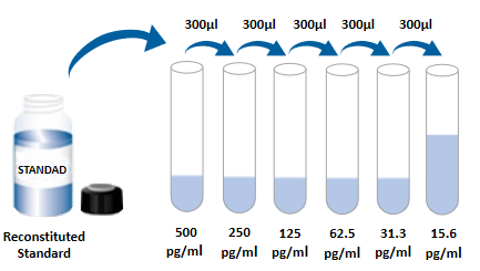 2. Operation steps 1. Prepare all required reagents and standards; 2. Take out the microplate from the sealed bag that has been equilibrated to room temperature. Please put the unused slats back into the aluminum foil bag and re-seal; 3. Add to the microplate 300uL Washing liquid, let stand and soak 30 Seconds, discard the lotion and pat the microplate dry on absorbent paper. Please use it immediately and do not let the microplate dry; 4. Add different concentration standards, experimental samples or quality control articles into the corresponding wells, and each well 100uL 。 Seal the reaction wells with plate sealing tape and incubate at room temperature 2 hour ; 5. Suck the liquid in the plate and wash the plate using a washing bottle, a multi-channel plate washer or an automatic plate washer. Washing solution per well 300uL Then the wash liquid in the plate is aspirated off. Repeat Operation 3 Times. Trying to absorb the residual liquid as much as possible every time you wash the plate will help to get good experimental results. At the end of the last plate washing, please suck all the liquid in the plate or turn the plate upside down, and pat all the residual liquid dry on absorbent paper; 6. Within each well added 100uL Detect antibodies. Seal the reaction wells with plate sealing tape and incubate at room temperature 2 Hour; 7. Repeat th 5 Step washing operation; 8. Within each well added 100uLSA-HRP , room temperature incubation 20 Minutes. Be careful to avoid light; 9. Repeat th 5 Step washing operation; 10. Within each well added 100uL Chromogenic solution, incubate at room temperature 5-30 Minutes, pay attention to avoiding light; 11. Within each well added 50uL Stop solution, the color of the solution in the well will change from blue to yellow. If the color of the solution changes to green or the color changes are inconsistent, pat the microplate gently to mix the solution evenly; 12. After addition of stop solution 30 Within minutes, measured using a plate reader 450nm Absorbance value, set 540nm Or 570nm As the correction wavelength. If dual-wavelength correction is not used, the accuracy of the results may be affected; 13. Calculation results: Add the corrected absorbance values of each standard and sample (OD450-OD540 Or OD570) , average of repeated well readings and then subtract the average zero standard OD Value. Using computer software for four-parameter logic (4-PL) Curve fitting creates a standard curve. Another way is to plot the standard concentration and make the logarithm with the corresponding OD The values were logarithmic to generate a curve, and the best fit line was determined by regression analysis. This process can generate a data fit that is sufficiently useful but less accurate. If the sample is diluted, the concentration should be calculated by multiplying the dilution factor.  3. Kit parameters 1. Recovery: Different levels of human were spiked in cell culture medium samples IL-2 The recovery rate was determined. The recoveries range from 102-104% , the average recovery was in 103% 。 2. Sensitivity: Human IL-2 The lowest measurable dose ( MDD ) is generally less than 3.24pg/mL 。 The lowest measurable value is determined according to 20 The corresponding concentration is calculated by adding two standard deviations to the mean value of the zero-point absorbance values of each standard curve. 3. Correction: This ELISA High purity recombinant human expressed by Escherichia coli IL-2 Corrected by protein. 4. Linearity: 4 Different samples were spiked with high concentrations of human IL-2 , followed by a diluent ( 1× ) Dilute the sample to the detection range and determine its linearity.
5. Specificity: This ELISA Method can detect natural and recombinant human IL-2 Egg whites. The following factors were mixed with diluent ( 1× ) formulated into 50ng/mL Concentration to detect human IL-2 Cross-reactivity of. Will 50ng/mL Interference factors incorporated into the intermediate range of recombinant human IL-2 In the reference substance, to detect the effects of human IL-2 Of interference. No significant cross-reactivity or interference was observed.
4. Analysis of frequently asked questions 1. Whiteboard (no color appears after color rendering is complete)
2. Flower plate (blank, negative positive control normal, but specimen well OD Values are significantly higher)
5. Experimental flow chart  |
||||||||||||||||||||||||||||||||||||||||||||||||||||||||||||||||||||
| Species Reactivity | Human | ||||||||||||||||||||||||||||||||||||||||||||||||||||||||||||||||||||
| Theory | This kit adopts double antibody sandwich enzyme-linked immunosorbent detection technology. Specific anti-human IL-2 antibodies were pre-coated on high affinity labeled plates. The standard substance, the sample to be tested and the biotinylated detection antibody are added to the well of the enzyme label plate, and after incubation, IL-2 present in the sample binds to the solid phase antibody and the detection antibody to form an immune complex. After washing to remove unbound material, horseradish peroxidase-labeled Streptavidin-HRP was added. After washing, a chromogenic substrate is added to protect the color from light. A stop solution was added to stop the reaction, and the absorbance value was measured at a wavelength of 450 nm (reference calibration wavelength of 540 nm or 570 nm). | ||||||||||||||||||||||||||||||||||||||||||||||||||||||||||||||||||||
| Synonym | aldesleukin, IL2, IL-2, IL-2lymphokine, interleukin 2, interleukin-2, involved in regulation of T-cell clonal expansion, T cell growth factor, T-cell growth factor, TCGF, Human interleukin 2 Elisa kit | ||||||||||||||||||||||||||||||||||||||||||||||||||||||||||||||||||||
| Composition |
Please use it within the validity period of the kit (new and old products are shipped randomly)
|
||||||||||||||||||||||||||||||||||||||||||||||||||||||||||||||||||||
| Background | Human interleukin 2 (IL-2), also known as T cell growth factor (TCGF), is an α-helix polypeptide with a molecular weight of 15-18kDa with different glycosylations, which belongs to the common γ-chain (γc) cytokine family members. It exists as a monomer with a very short half-life (< 30min). The precursor of human IL-2 has 153 amino acids, including a 20 amino acid signal sequence, and a 133 amino acid mature polypeptide. IL-2 mature protein contains an O-type glycosylation site at threonine position 3 and three cysteines, of which the intra-chain disulfide bond formed by two cysteines is essential for IL-2 activity. The mature human IL-2 amino acid sequence shares 73%, 66%, 78%, and 97% homology to IL-2 in dog, rat, cat, and macaque, respectively. Although human IL-2 has only 60% amino acid homology with highly polymorphic mouse IL-2, human IL-2 is also biologically active on mouse cells. Cells reported to secrete IL-2 include γδ T cells, activated conventional CD4 + and CD8 + T cells, neurons, microglia, hematopoietic stem cells. The IL-2 receptor (IL-2R) consists of three subunits, a 55 kDa CD25/IL-2Rα chain, a 70 kDa CD122/IL-2Rβ chain, and a 65 kDa CD132/γc chain. IL-2 first binds to CD25, and the two-subunit complex formed recruits CD122 and CD132, forming a signal complex with four subunits. In addition to forming a complex with IL-2, CD122/IL-2Rβ can also form a four-subunit signaling complex with IL-15. CD132/ In vitro studies have shown that IL-2 plays an important role in T cell activation and expansion. In vivo, IL-2 is key to the development, maintenance, and function of regulatory T cells (Tregs), and regulatory T cells can provide protection against autoimmune diseases. In addition, IL-2 can also promote autoimmune inflammation in its target organs by regulating the expression of T cell trafficking genes and the production of Th2 cytokines. In CD8 + T cell subsets, IL-2 is a necessary key to obtaining an optimal first-order response and differentiation into terminal effector cells. IL-2 also promotes the development and activation of CD8 + T cells into memory cells. |
||||||||||||||||||||||||||||||||||||||||||||||||||||||||||||||||||||
| General Notes | 1. Please use the kit within the validity period. 2. The components of different kits and different batch kits cannot be mixed. 3. If the sample value is greater than the highest value of the standard curve, the sample should be diluted with diluent (1 ×) and re-tested; If the cell culture supernatant sample needs to be distributed and diluted, cell culture medium can be used for other intermediate dilutions except dilution with diluent in the last step. 4. Differences in test results can be caused by a variety of factors, including the operation of the experimenter, the use of the pipette, the plate washing technique, the reaction time or temperature, the storage of the kit, etc. 5. The terminating solution in the kit is an acidic solution. Please protect your glasses, hands, face and clothes when using it. 6. For scientific research only, not for in vitro diagnosis. |
||||||||||||||||||||||||||||||||||||||||||||||||||||||||||||||||||||
| Storage Temp. | Kit unopened, stored at 2-8 ℃. | ||||||||||||||||||||||||||||||||||||||||||||||||||||||||||||||||||||
| Test Range | 15.6pg/mL-1000pg/mL |
Picture
Picture
Immunohistochemistry