WB result of TBK1/NAK Rabbit mAb
Primary antibody: TBK1/NAK Rabbit mAb at 1/1000 dilution
Lane 1: HCT 116 whole cell lysate 20 µg
Lane 2: HeLa whole cell lysate 20 µg
Lane 3: MCF7 whole cell lysate 20 µg
Lane 4: SH-SY5Y whole cell lysate 20 µg
Secondary antibody: Goat Anti-Rabbit IgG, (H+L), HRP conjugated at 1/10000 dilution
Predicted MW: 84 kDa
Observed MW: 84 kDa
(This blot was developed with high sensitivity substrate)
TBK1/NAK Recombinant Rabbit mAb (S-658-50)
TBK1/NAK Recombinant Rabbit mAb (S-658-50)
Price:
Regular price
$100 USD
Regular price
Sale price
$100 USD
Unit price
per
For shipping services or bulk orders, you may request a quotation.
Secure checkout with

View full details
Product Details
Product Details
Product Specification
| Host | Rabbit |
| Antigen | TBK1/NAK |
| Synonyms | Serine/threonine-protein kinase TBK1, NF-kappa-B-activating kinase, T2K, TANK-binding kinase 1 |
| Immunogen | Synthetic Peptide |
| Location | Cytoplasm |
| Accession | Q9UHD2 |
| Clone Number | S-658-50 |
| Antibody Type | Recombinant mAb |
| Isotype | IgG |
| Application | WB, IHC-P, ICC, ICFCM |
| Reactivity | Hu, Ms, Rt |
| Purification | Protein A |
| Concentration | 2 mg/ml |
| Conjugation | Unconjugated |
| Physical Appearance | Liquid |
| Storage Buffer | PBS, 40% Glycerol, 0.05%BSA, 0.03% Proclin 300 |
| Stability & Storage | 12 months from date of receipt / reconstitution, -20 °C as supplied |
Dilution
| application | dilution | species |
| WB | 1:1000 | |
| IHC-P | 1:200 | |
| ICFCM | 1:200 | |
| ICC | 1:400 |
Background
TBK1 (TANK-binding kinase 1) is an enzyme with kinase activity. Specifically, it is a serine / threonine protein kinase. This kinase is mainly known for its role in innate immunity antiviral response. However, TBK1 also regulates cell proliferation, apoptosis, autophagy, and anti-tumor immunity. Insufficient regulation of TBK1 activity leads to autoimmune, neurodegenerative diseases or tumorigenesis. As a non-canonical IKK, TBK1 is also involved in the non-canonical NFkB pathway. It phosphorylates p100/NF-κB2, which is subsequently processed in the proteasome and released as a p52 subunit. This subunit dimerizes with RelB and mediates gene expression.
Picture
Picture
Western Blot
WB result of TBK1/NAK Rabbit mAb
Primary antibody: TBK1/NAK Rabbit mAb at 1/1000 dilution
Lane 1: NIH/3T3 whole cell lysate 20 µg
Secondary antibody: Goat Anti-Rabbit IgG, (H+L), HRP conjugated at 1/10000 dilution
Predicted MW: 84 kDa
Observed MW: 84 kDa
WB result of TBK1/NAK Rabbit mAb
Primary antibody: TBK1/NAK Rabbit mAb at 1/1000 dilution
Lane 1: C6 whole cell lysate 20 µg
Lane 2: rat testis lysate 20 µg
Secondary antibody: Goat Anti-Rabbit IgG, (H+L), HRP conjugated at 1/10000 dilution
Predicted MW: 84 kDa
Observed MW: 84 kDa
FC
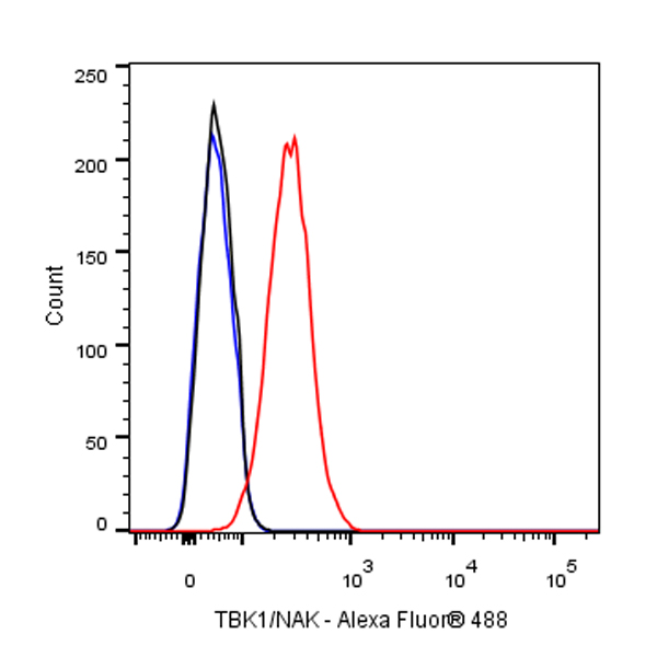
Flow cytometric analysis of 4% PFA fixed 90% methanol permeabilized MCF7 (Human breast adenocarcinoma epithelial cell) cells labelling TBK1/NAK antibody at 1/200 dilution (1 μg)/ (Red) compared with a Rabbit monoclonal IgG (Black) isotype control and an unlabelled control (cells without incubation with primary antibody and secondary antibody) (Blue). Goat Anti - Rabbit IgG Alexa Fluor® 488 was used as the secondary antibody.
Immunohistochemistry
IHC shows positive staining in paraffin-embedded human stomach. Anti-TBK1/NAK antibody was used at 1/200 dilution, followed by a HRP Polymer for Mouse & Rabbit IgG (ready to use). Counterstained with hematoxylin. Heat mediated antigen retrieval with Tris/EDTA buffer pH9.0 was performed before commencing with IHC staining protocol.
IHC shows positive staining in paraffin-embedded mouse testis. Anti-TBK1/NAK antibody was used at 1/200 dilution, followed by a HRP Polymer for Mouse & Rabbit IgG (ready to use). Counterstained with hematoxylin. Heat mediated antigen retrieval with Tris/EDTA buffer pH9.0 was performed before commencing with IHC staining protocol.
IHC shows positive staining in paraffin-embedded rat testis. Anti-TBK1/NAK antibody was used at 1/200 dilution, followed by a HRP Polymer for Mouse & Rabbit IgG (ready to use). Counterstained with hematoxylin. Heat mediated antigen retrieval with Tris/EDTA buffer pH9.0 was performed before commencing with IHC staining protocol.
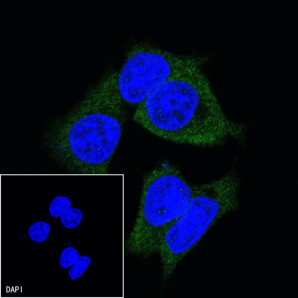
Immunocytochemistry
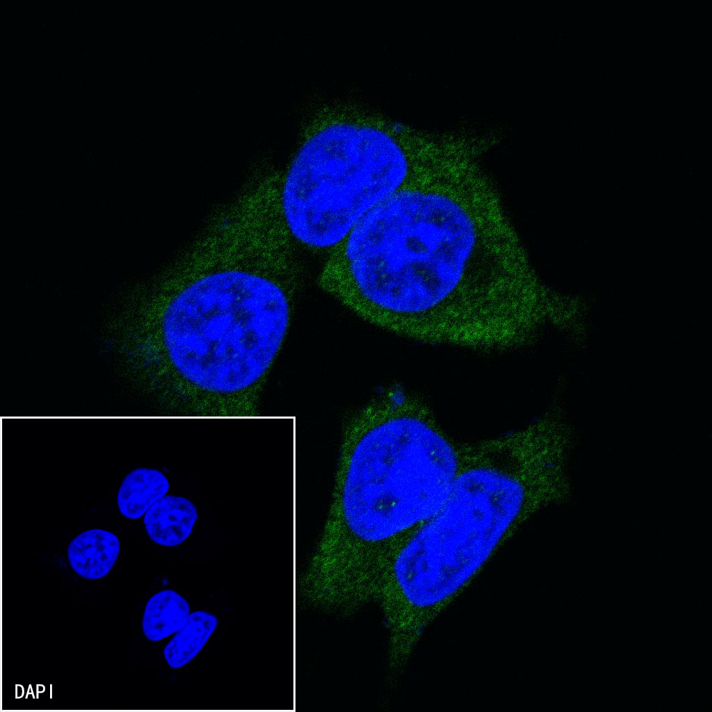
ICC shows positive staining in MCF7 cells. Anti-TBK1/NAK antibody was used at 1/400 dilution (Green) and incubated overnight at 4°C. Goat polyclonal Antibody to Rabbit IgG - H&L (Alexa Fluor® 488) was used as secondary antibody at 1/1000 dilution. The cells were fixed with 100% ice-cold methanol and permeabilized with 0.1% PBS-Triton X-100. Nuclei were counterstained with DAPI (Blue).