WB result of SLC7A5/LAT1 Rabbit mAb Primary antibody: SLC7A5/LAT1 Rabbit mAb at 1/1000 dilution Lane 1: LNCaP whole cell lysate 20 µg Lane 2: HT-29 whole cell lysate 20 µg Lane 3: HT-1080 whole cell lysate 20 µg Lane 4: MCF7 whole cell lysate 20 µg Negative control: LNCaP whole cell lysate Secondary antibody: Goat Anti-Rabbit IgG, (H+L), HRP conjugated at 1/10000 dilution Predicted MW: 55 kDa Observed MW: 36 kDa
SLC7A5/LAT1 Recombinant Rabbit mAb (S-367-12)
SLC7A5/LAT1 Recombinant Rabbit mAb (S-367-12)
Price:
Regular price
$100 USD
Regular price
Sale price
$100 USD
Unit price
per
For shipping services or bulk orders, you may request a quotation.
Secure checkout with

View full details
Product Details
Product Details
Product Specification
| Host | Rabbit |
| Antigen | SLC7A5/LAT1 |
| Synonyms | Large neutral amino acids transporter small subunit 1, 4F2 light chain (4F2 LC; 4F2LC), CD98LC, MPE16, hLAT1, y+ system cationic amino acid transporter |
| Immunogen | Synthetic Peptide |
| Location | Membrane, Lysosome |
| Accession | Q01650 |
| Clone Number | S-367-12 |
| Antibody Type | Rabbit mAb |
| Application | WB, IHC-P, ICC |
| Reactivity | Hu |
| Purification | Protein A |
| Concentration | 0.5 mg/ml |
| Conjugation | Unconjugated |
| Physical Appearance | Liquid |
| Storage Buffer | PBS, 40% Glycerol, 0.05% BSA, 0.03% Proclin 300 |
| Stability & Storage | 12 months from date of receipt / reconstitution, -20 °C as supplied |
Dilution
| application | dilution | species |
| WB | 1:1000 | |
| IHC | 1:2000 | |
| ICC | 1:500 |
Background
SLC7A5 (Large neutral amino acids transporter small subunit 1), also known as 4F2 light chain, or CD98 light chain is a protein that in humans is encoded by the SLC7A5 gene. It is suggested to play a part in altered cell metabolism and proliferative signaling and has been reported to be overexpressed in various types of cancer, including breast cancer [PMID: 34792178].
Picture
Picture
Western Blot
Immunohistochemistry
IHC shows positive staining in paraffin-embedded human cerebral cortex. Anti-SLC7A5/LAT1 antibody was used at 1/2000 dilution, followed by a HRP Polymer for Mouse & Rabbit IgG (ready to use). Counterstained with hematoxylin. Heat mediated antigen retrieval with Tris/EDTA buffer pH9.0 was performed before commencing with IHC staining protocol.
IHC shows positive staining in paraffin-embedded human testis. Anti-SLC7A5/LAT1 antibody was used at 1/2000 dilution, followed by a HRP Polymer for Mouse & Rabbit IgG (ready to use). Counterstained with hematoxylin. Heat mediated antigen retrieval with Tris/EDTA buffer pH9.0 was performed before commencing with IHC staining protocol.
IHC shows positive staining in paraffin-embedded human breast cancer. Anti-SLC7A5/LAT1 antibody was used at 1/2000 dilution, followed by a HRP Polymer for Mouse & Rabbit IgG (ready to use). Counterstained with hematoxylin. Heat mediated antigen retrieval with Tris/EDTA buffer pH9.0 was performed before commencing with IHC staining protocol.
IHC shows positive staining in paraffin-embedded human lung cancer. Anti-SLC7A5/LAT1 antibody was used at 1/2000 dilution, followed by a HRP Polymer for Mouse & Rabbit IgG (ready to use). Counterstained with hematoxylin. Heat mediated antigen retrieval with Tris/EDTA buffer pH9.0 was performed before commencing with IHC staining protocol.
IHC shows positive staining in paraffin-embedded human cervical squamous cell carcinoma. Anti-SLC7A5/LAT1 antibody was used at 1/2000 dilution, followed by a HRP Polymer for Mouse & Rabbit IgG (ready to use). Counterstained with hematoxylin. Heat mediated antigen retrieval with Tris/EDTA buffer pH9.0 was performed before commencing with IHC staining protocol.
Immunocytochemistry
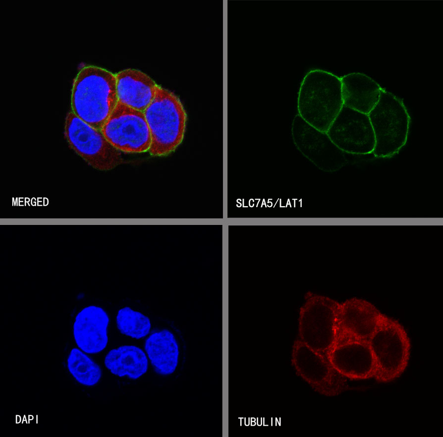
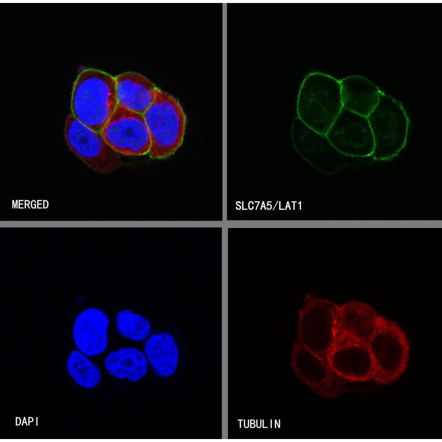
ICC shows positive staining in MCF7 cells. Anti-SLC7A5/LAT1 antibody was used at 1/500 dilution (Green) and incubated overnight at 4°C. Goat polyclonal Antibody to Rabbit IgG - H&L (Alexa Fluor® 488) was used as secondary antibody at 1/1000 dilution. The cells were fixed with 4%PFA and permeabilized with 0.1% PBS-Triton X-100. Nuclei were counterstained with DAPI (Blue).Counterstain with tubulin (Red).
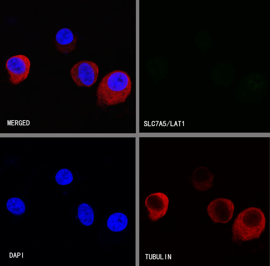
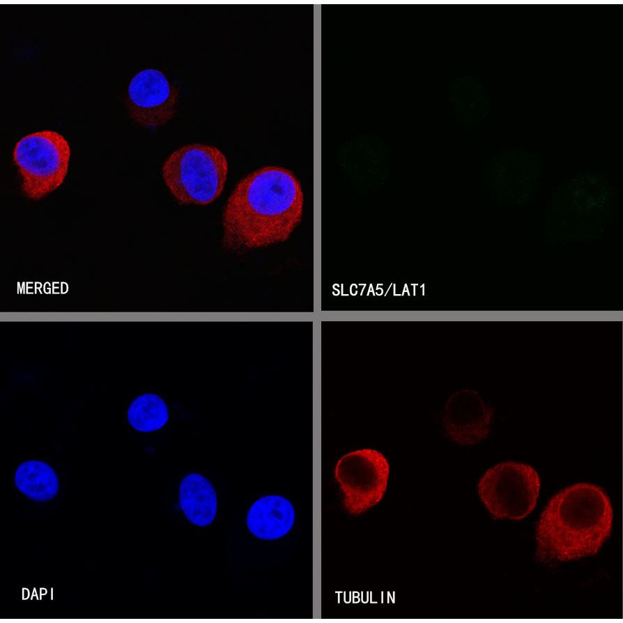
Negative control:ICC shows negative staining in LNCAP cells. Anti-SLC7A5/LAT1 antibody was used at 1/500 dilution and incubated overnight at 4°C. Goat polyclonal Antibody to Rabbit IgG - H&L (Alexa Fluor® 488) was used as secondary antibody at 1/1000 dilution. The cells were fixed with 4%PFA and permeabilized with 0.1% PBS-Triton X-100. Nuclei were counterstained with DAPI (Blue).Counterstain with tubulin (red).