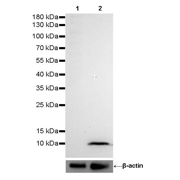
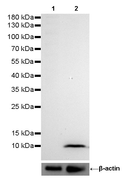
WB result of S100A8 (MRP8) Rabbit mAb
Primary antibody: S100A8 (MRP8) Rabbit mAb at 1/1000 dilution
Lane 1: 293T whole cell lysate 20 µg
Lane 2: SK-BR-3 whole cell lysate 20 µg
Negative control: 293T whole cell lysate
Secondary antibody: Goat Anti-Rabbit IgG, (H+L), HRP conjugated at 1/10000 dilution
Predicted MW: 10 kDa
Observed MW: 10 kDa
Exposure time: 10s
S100A8 (MRP8) Recombinant Rabbit mAb (SDT-059-28)
S100A8 (MRP8) Recombinant Rabbit mAb (SDT-059-28)
Price:
Regular price
$100 USD
Regular price
Sale price
$100 USD
Unit price
per
For shipping services or bulk orders, you may request a quotation.
Secure checkout with

View full details
Product Details
Product Details
Product Specification
| Host | Rabbit |
| Antigen | S100A8 (MRP8) |
| Synonyms | Protein S100-A8, CFAG |
| Immunogen | Synthetic Peptide |
| Location | Cytoplasm, Secreted, Cell membrane |
| Accession | P05109 |
| Clone Number | SDT-059-28 |
| Antibody Type | Rabbit mAb |
| Application | WB, IHC-P, ICC, IP |
| Reactivity | Hu, Ms, Rt |
| Purification | Protein A |
| Concentration | 0.5 mg/ml |
| Physical Appearance | Liquid |
| Storage Buffer | PBS, 40% Glycerol, 0.05 %BSA, 0.03% Proclin 300 |
| Stability & Storage | 12 months from date of receipt / reconstitution, -20 °C as supplied |
Dilution
| application | dilution | species |
| WB | 1:1000 | |
| IHC-P | 1:2000 | |
| IP | 1:50 | |
| ICC | 1:500 |
Background
S100A8 (MRP8) is a calcium- and zinc-binding protein which plays a prominent role in the regulation of inflammatory processes and immune response. It can induce neutrophil chemotaxis and adhesion. Predominantly found as calprotectin (S100A8/A9) which has a wide plethora of intra- and extracellular functions. The intracellular functions include: facilitating leukocyte arachidonic acid trafficking and metabolism, modulation of the tubulin-dependent cytoskeleton during migration of phagocytes and activation of the neutrophilic NADPH-oxidase.
Picture
Picture
Western Blot
IP
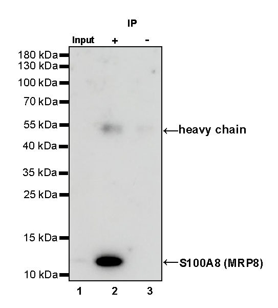
S100A8 (MRP8) Rabbit mAb at 1/50 dilution (1 µg) immunoprecipitating S100A8 (MRP8) in 0.4 mg HL-60 whole cell lysate.
Western blot was performed on the immunoprecipitate using S100A8 (MRP8) Rabbit mAb at 1/1000 dilution.
Secondary antibody (HRP) for IP was used at 1/400 dilution.
Lane 1: HL-60 whole cell lysate 20 µg (Input)
Lane 2: S100A8 (MRP8) Rabbit mAb IP in HL-60 whole cell lysate
Lane 3: Rabbit monoclonal IgG IP in HL-60 whole cell lysate
Predicted MW: 10 kDa
Observed MW: 12 kDa
Exposure time: 39 s
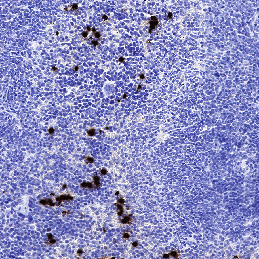
Immunohistochemistry

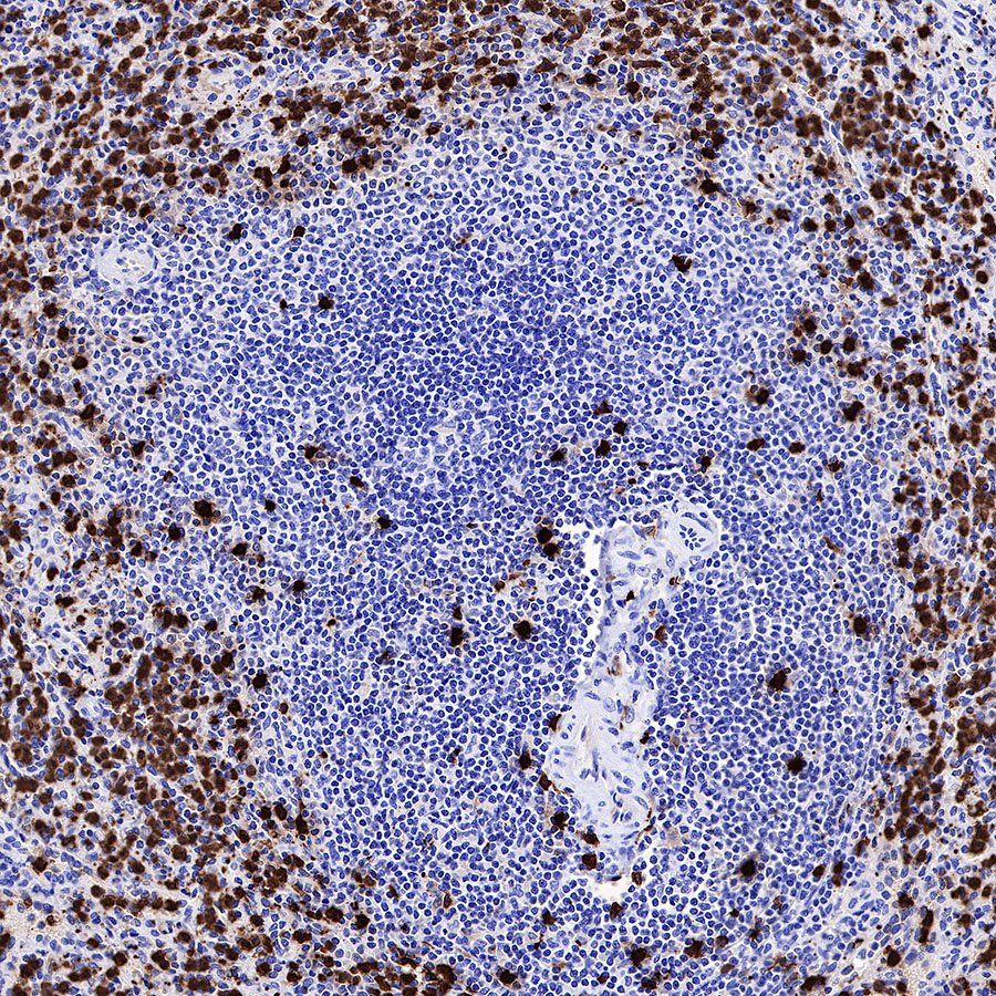
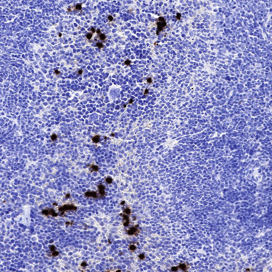
IHC shows positive staining in paraffin-embedded human spleen. Anti-S100A8 (MRP8) antibody was used at 1/2000 dilution, followed by a HRP Polymer for Mouse & Rabbit IgG (ready to use). Counterstained with hematoxylin. Heat mediated antigen retrieval with Tris/EDTA buffer pH9.0 was performed before commencing with IHC staining protocol.

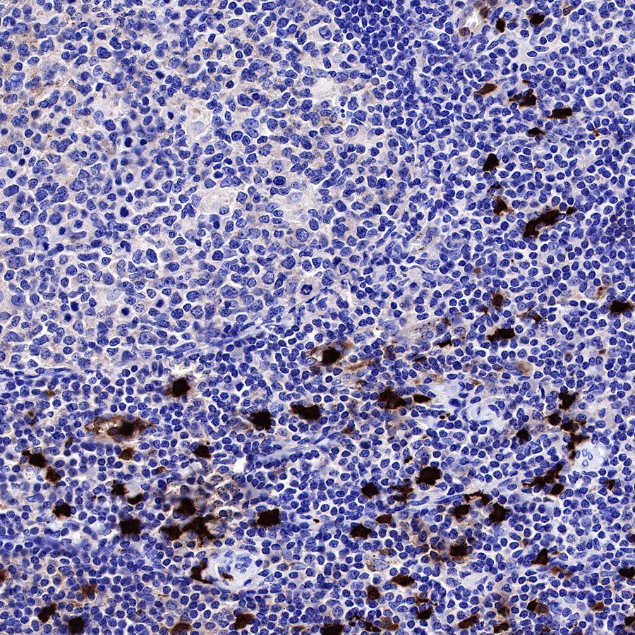
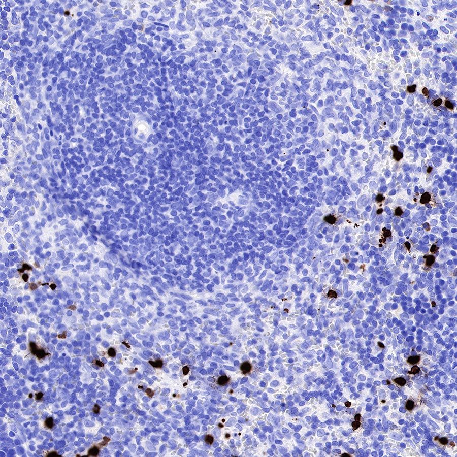
IHC shows positive staining in paraffin-embedded human tonsil. Anti-S100A8 (MRP8) antibody was used at 1/2000 dilution, followed by a HRP Polymer for Mouse & Rabbit IgG (ready to use). Counterstained with hematoxylin. Heat mediated antigen retrieval with Tris/EDTA buffer pH9.0 was performed before commencing with IHC staining protocol.

IHC shows positive staining in paraffin-embedded human tonsil. Anti-S100A8 (MRP8) antibody was used at 1/2000 dilution, followed by a HRP Polymer for Mouse & Rabbit IgG (ready to use). Counterstained with hematoxylin. Heat mediated antigen retrieval with Tris/EDTA buffer pH9.0 was performed before commencing with IHC staining protocol.

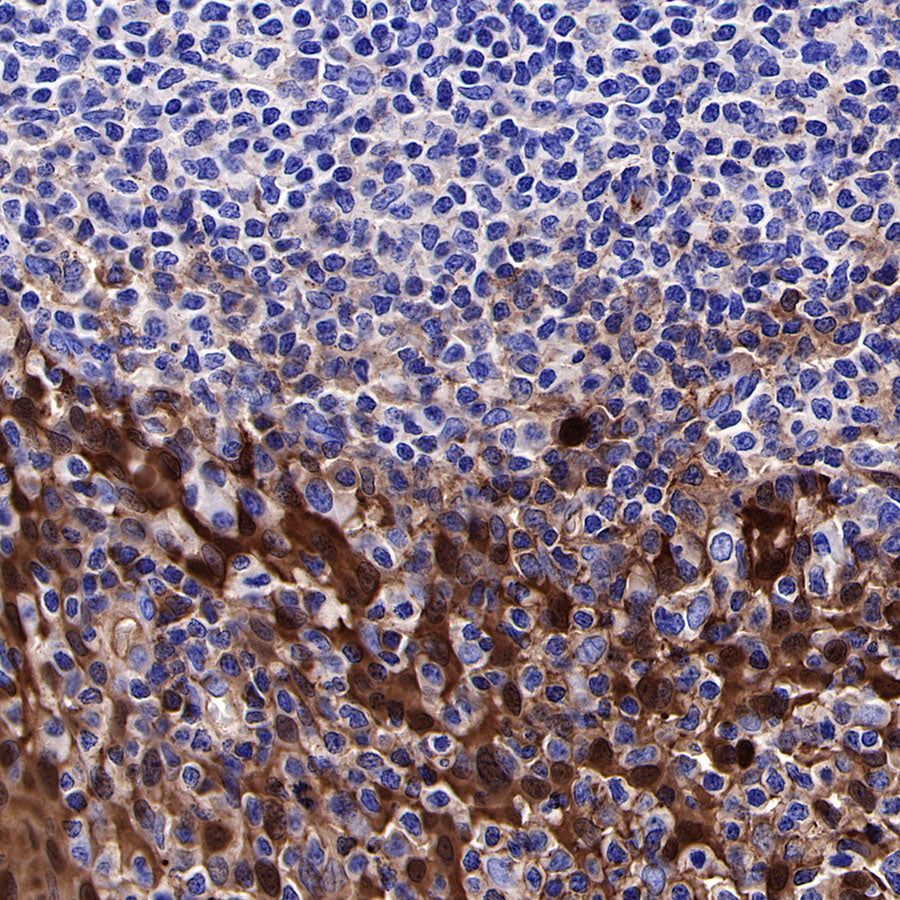
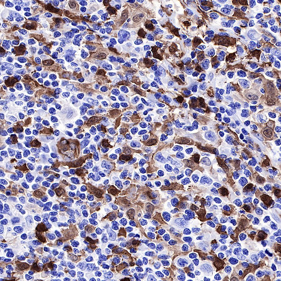
IHC shows positive staining in paraffin-embedded human Hodgkin's lymphoma. Anti-S100A8 (MRP8) antibody was used at 1/2000 dilution, followed by a HRP Polymer for Mouse & Rabbit IgG (ready to use). Counterstained with hematoxylin. Heat mediated antigen retrieval with Tris/EDTA buffer pH9.0 was performed before commencing with IHC staining protocol.
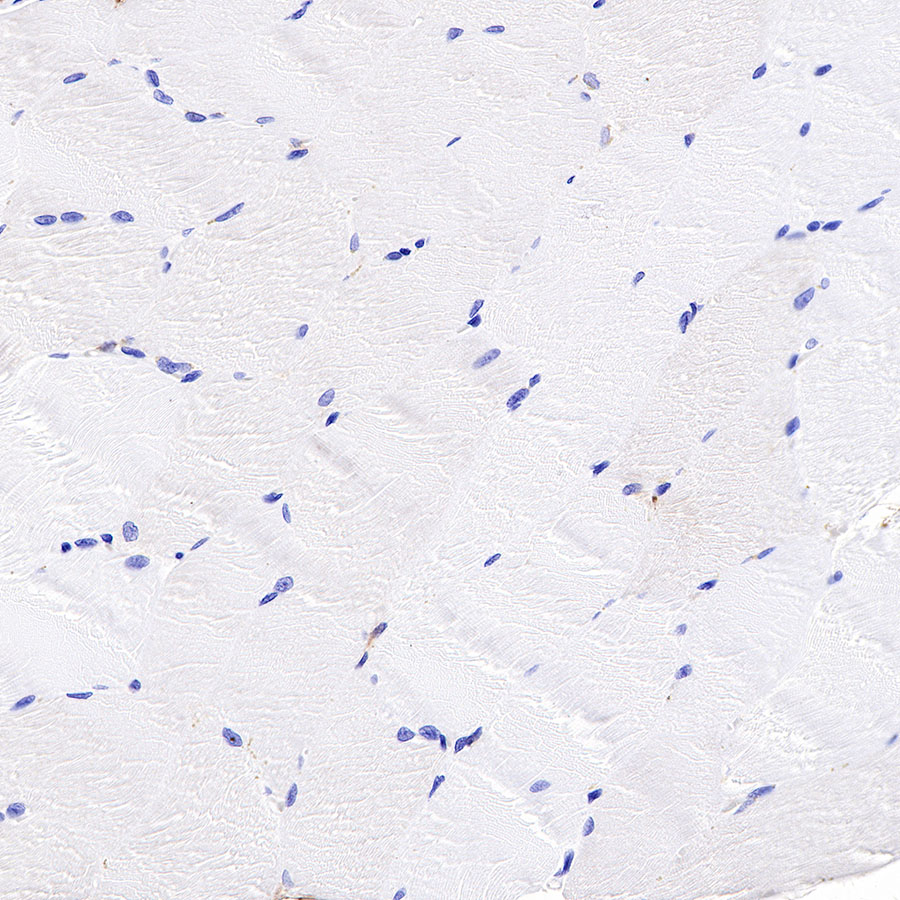
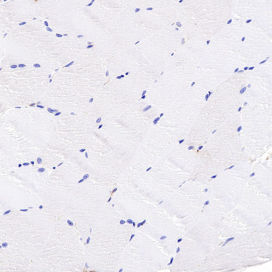
Negative control: IHC shows negative staining in paraffin-embedded human skeletal muscle. Anti-S100A8 (MRP8) antibody was used at 1/2000 dilution, followed by a HRP Polymer for Mouse & Rabbit IgG (ready to use). Counterstained with hematoxylin. Heat mediated antigen retrieval with Tris/EDTA buffer pH9.0 was performed before commencing with IHC staining protocol.
IHC shows positive staining in paraffin-embedded mouse spleen. Anti-S100A8 (MRP8) antibody was used at 1/2000 dilution, followed by a HRP Polymer for Mouse & Rabbit IgG (ready to use). Counterstained with hematoxylin. Heat mediated antigen retrieval with Tris/EDTA buffer pH9.0 was performed before commencing with IHC staining protocol.

IHC shows positive staining in paraffin-embedded rat spleen. Anti-S100A8 (MRP8) antibody was used at 1/2000 dilution, followed by a HRP Polymer for Mouse & Rabbit IgG (ready to use). Counterstained with hematoxylin. Heat mediated antigen retrieval with Tris/EDTA buffer pH9.0 was performed before commencing with IHC staining protocol.
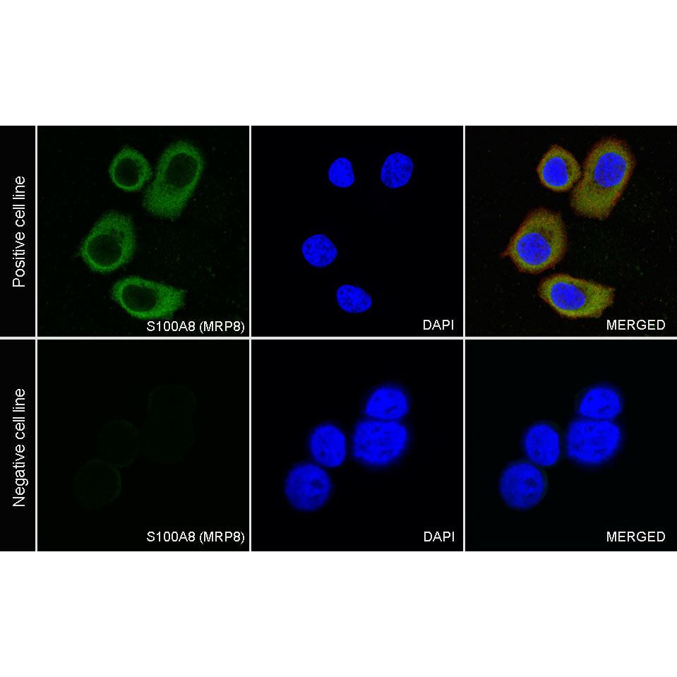
Immunocytochemistry
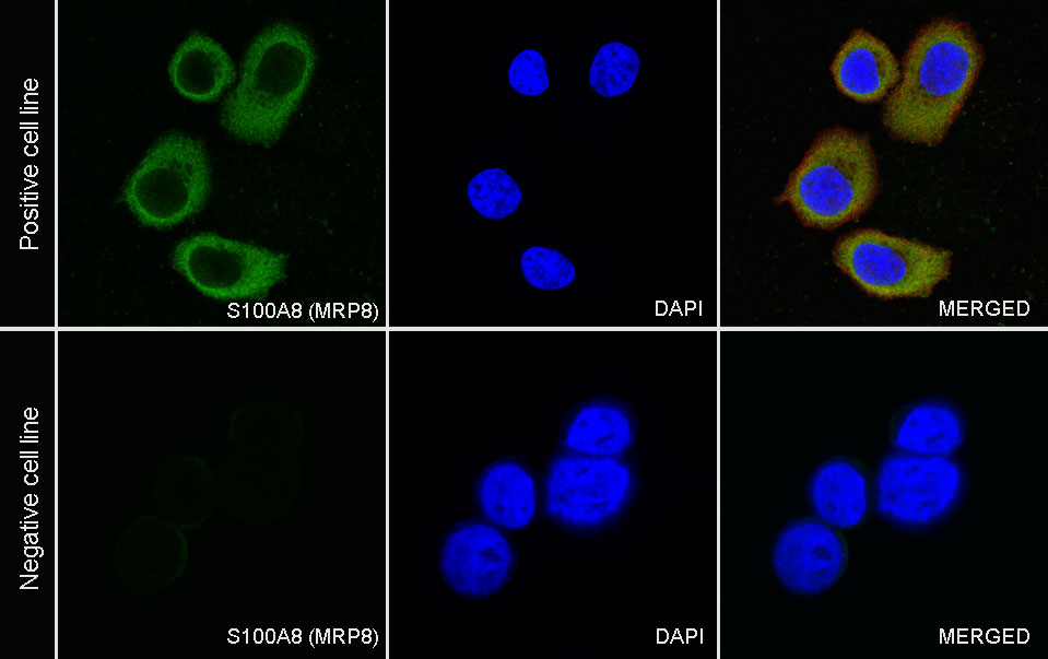
ICC shows positive staining in SK-BR-3 cells (top panel) and negative staining in 293T cells (below panel). Anti-S100A8 (MRP8) antibody was used at 1/500 dilution (Green) and incubated overnight at 4°C. Goat polyclonal Antibody to Rabbit IgG - H&L (Alexa Fluor® 488) was used as secondary antibody at 1/1000 dilution. The cells were fixed with 100% ice-cold methanol and permeabilized with 0.1% PBS-Triton X-100. Nuclei were counterstained with DAPI (Blue). Counterstain with tubulin (Red).