WB result of RPL26L1 Rabbit mAb
Primary antibody: RPL26L1 Rabbit mAb at 1/1000 dilution
Lane 1: 293T whole cell lysate 5 µg
Lane 2: HepG2 whole cell lysate 5 µg
Lane 3: HeLa whole cell lysate 5 µg
Secondary antibody: Goat Anti-Rabbit IgG, (H+L), HRP conjugated at 1/10000 dilution
Predicted MW: 17 kDa
Observed MW: 17 kDa
RPL26L1 Recombinant Rabbit mAb (S-R317)
RPL26L1 Recombinant Rabbit mAb (S-R317)
Price:
Regular price
$100 USD
Regular price
Sale price
$100 USD
Unit price
per
For shipping services or bulk orders, you may request a quotation.
Secure checkout with

View full details
Product Details
Product Details
Product Specification
| Host | Rabbit |
| Synonyms | Ribosomal protein uL24-like, 60S ribosomal protein L26-like 1, Large ribosomal subunit protein uL24-like 1, RPL26P1 |
| Accession | Q9UNX3 |
| Clone Number | S-R317 |
| Antibody Type | Recombinant mAb |
| Isotype | IgG |
| Application | WB, IHC-P, IP |
| Reactivity | Hu |
| Purification | Protein A |
| Concentration | 0.5 mg/ml |
| Conjugation | Unconjugated |
| Physical Appearance | Liquid |
| Storage Buffer | PBS, 40% Glycerol, 0.05% BSA, 0.03% Proclin 300 |
| Stability & Storage | 12 months from date of receipt / reconstitution, -20 °C as supplied |
Dilution
| application | dilution | species |
| WB | 1:1000 | null |
| IHC-P | 1:1000 | null |
| IP | 1:50 | null |
Background
RPL26L1 is a protein that shares high sequence similarity with RPL26. It is not currently known whether the protein is a functional ribosomal protein or whether it has evolved a function that is independent of the ribosome.
Picture
Picture
Western Blot
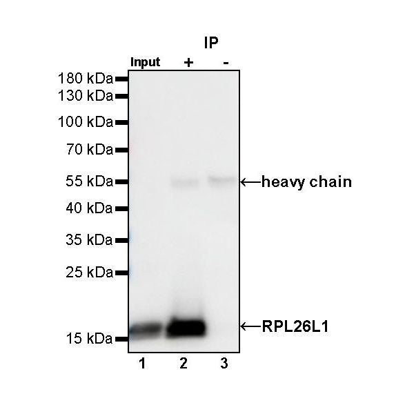
IP
RPL26L1 Rabbit mAb at 1/50 dilution (1 µg) immunoprecipitating RPL26L1 in 0.4 mg HeLa whole cell lysate.
Western blot was performed on the immunoprecipitate using RPL26L1 Rabbit mAb at 1/1000 dilution.
Secondary antibody (HRP) for IP was used at 1/400 dilution.
Lane 1: HeLa whole cell lysate 20 µg (Input)
Lane 2: RPL26L1 Rabbit mAb IP in HeLa whole cell lysate
Lane 3: Rabbit monoclonal IgG IP in HeLa whole cell lysate
Predicted MW: 17 kDa
Observed MW: 17 kDa
Immunohistochemistry
IHC shows positive staining in paraffin-embedded human colon. Anti-RPL26L1 antibody was used at 1/1000 dilution, followed by a HRP Polymer for Mouse & Rabbit IgG (ready to use). Counterstained with hematoxylin. Heat mediated antigen retrieval with Tris/EDTA buffer pH9.0 was performed before commencing with IHC staining protocol.
IHC shows positive staining in paraffin-embedded human kidney. Anti-RPL26L1 antibody was used at 1/1000 dilution, followed by a HRP Polymer for Mouse & Rabbit IgG (ready to use). Counterstained with hematoxylin. Heat mediated antigen retrieval with Tris/EDTA buffer pH9.0 was performed before commencing with IHC staining protocol.
IHC shows positive staining in paraffin-embedded human pancreas. Anti-RPL26L1 antibody was used at 1/1000 dilution, followed by a HRP Polymer for Mouse & Rabbit IgG (ready to use). Counterstained with hematoxylin. Heat mediated antigen retrieval with Tris/EDTA buffer pH9.0 was performed before commencing with IHC staining protocol.
Negative control: IHC shows negative staining in paraffin-embedded human skeletal muscle. Anti-RPL26L1 antibody was used at 1/1000 dilution, followed by a HRP Polymer for Mouse & Rabbit IgG (ready to use). Counterstained with hematoxylin. Heat mediated antigen retrieval with Tris/EDTA buffer pH9.0 was performed before commencing with IHC staining protocol.
IHC shows positive staining in paraffin-embedded human lung squamous cell carcinoma. Anti-RPL26L1 antibody was used at 1/1000 dilution, followed by a HRP Polymer for Mouse & Rabbit IgG (ready to use). Counterstained with hematoxylin. Heat mediated antigen retrieval with Tris/EDTA buffer pH9.0 was performed before commencing with IHC staining protocol.