WB result of Phospho-IκBα (Ser36) Rabbit mAb Primary antibody: Phospho-IκBα (Ser36) Rabbit mAb at 1/1000 dilution Lane 1: untreated HeLa whole cell lysate 20 µg Lane 2: HeLa treated with Calyculin A (100 ng/ml, 30 min) +TNF-α (20 ng/ml, 5 min) whole cell lysate 20 µg Secondary antibody: Goat Anti-Rabbit IgG, (H+L), HRP conjugated at 1/10000 dilution Predicted MW: 35 kDa Observed MW: 39 kDa
Phospho-IκBα (Ser36) Recombinant Rabbit mAb (S-R102)
Phospho-IκBα (Ser36) Recombinant Rabbit mAb (S-R102)
Price:
Regular price
$100 USD
Regular price
Sale price
$100 USD
Unit price
per
For shipping services or bulk orders, you may request a quotation.
Secure checkout with

View full details
Product Details
Product Details
Product Specification
| Host | Rabbit |
| Antigen | Phospho-IκBα (Ser36) |
| Synonyms | Phospho-NF-kappa-B inhibitor alpha+ (Ser36), Phospho-I-kappa-B-alpha (Ser36), Phospho-IkB-alpha (Ser36), Phospho-IkappaBalpha (Ser36), Phospho-IKBA (Ser36), Phospho-MAD3 (Ser36), Phospho-NFKBIA (Ser36) |
| Location | Cytoplasm, Nucleus |
| Accession | P25963 |
| Clone Number | S-R102 |
| Antibody Type | Rabbit mAb |
| Application | WB, IHC-P, ICC, ICFCM |
| Reactivity | Hu |
| Purification | Protein A |
| Concentration | 0.5 mg/ml |
| Conjugation | Unconjugated |
| Physical Appearance | Liquid |
| Storage Buffer | PBS, 40% Glycerol, 0.05% BSA, 0.03% Proclin 300 |
| Stability & Storage | 12 months from date of receipt / reconstitution, -20 °C as supplied |
Dilution
| application | dilution | species |
| WB | 1:1000 | null |
| IHC | 1:2000 | null |
| ICFCM | 1:5000 | null |
| ICC | 1:500 | null |
Background
The IκBα (inhibitor of nuclear factor kappa B) protein inactivates the NF-κB transcription factor by masking the nuclear localization signals (NLS) of NF-κB proteins and keeping them sequestered in an inactive state in the cytoplasm [PMID: 9865693, PMID: 9244310, PMID: 9346484]. Activation occurs via phosphorylation of IκBα at Ser32 and Ser36 followed by proteasome-mediated degradation that results in the release and nuclear translocation of active NF-κB [PMID: 7739562]. Specifically, IKK phosphorylates the inhibitory IκBα protein [PMID: 10602462]. This phosphorylation results in the dissociation of IκBα from NF-κB. NF-κB, which is now free, migrates into the nucleus and activates the expression of at least 150 genes; some of which are anti-apoptotic.
Picture
Picture
Western Blot
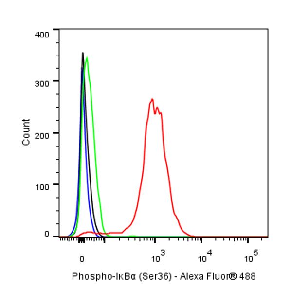
FC
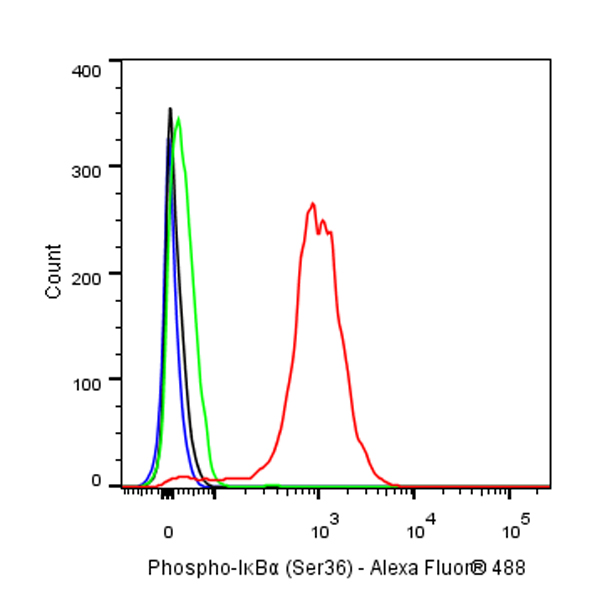
Flow cytometric analysis of HeLa (Human cervix adenocarcinoma epithelial cell) cells, treated with 100ng/ml Calyculin A for 30min, 20ng/ml TNF-α for 5min (Red) or untreated (Green), labeling Phospho-IκBα (Ser36) at 1/5000 dilution (0.01 μg) compared with a Rabbit monoclonal IgG isotype control (Black) and an unlabeled control (cells without incubation with primary antibody and secondary antibody) (Blue). Goat Anti - Rabbit IgG Alexa Fluor® 488 was used as the secondary antibody.
Immunohistochemistry
IHC shows positive staining in paraffin-embedded human cervical squamous cell carcinoma. Anti-Phospho-IκBα (Ser36) antibody was used at 1/2000 dilution, followed by a HRP Polymer for Mouse & Rabbit IgG (ready to use). Counterstained with hematoxylin. Heat mediated antigen retrieval with Tris/EDTA buffer pH9.0 was performed before commencing with IHC staining protocol.
IHC shows positive staining in paraffin-embedded human hepatocellular carcinoma. Anti-Phospho-IκBα (Ser36) antibody was used at 1/2000 dilution, followed by a HRP Polymer for Mouse & Rabbit IgG (ready to use). Counterstained with hematoxylin. Heat mediated antigen retrieval with Tris/EDTA buffer pH9.0 was performed before commencing with IHC staining protocol.
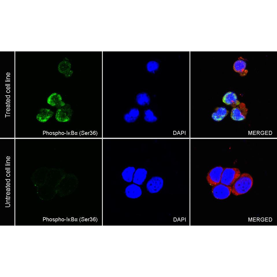
Immunocytochemistry
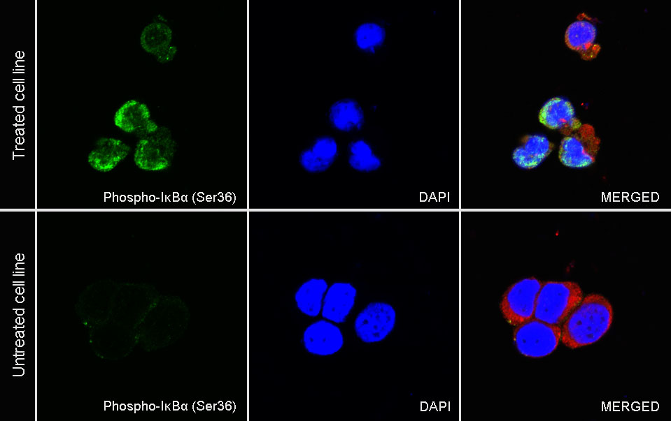
ICC analysis of HeLa cells treated with Calyculin A (100ng/ml, 30min)+TNF-α (20ng/ml, 5min) (top panel) and HeLa cells untreated with Calyculin A (100ng/ml, 30min)+TNF-α (20ng/ml, 5min) (below panel). Anti-Phospho-IκBα (Ser36) antibody was used at 1/500 dilution (Green) and incubated overnight at 4°C. Goat polyclonal Antibody to Rabbit IgG - H&L (Alexa Fluor® 488) was used as secondary antibody at 1/1000 dilution. The cells were fixed with 100% ice-cold methanol and permeabilized with 0.1% PBS-Triton X-100. Nuclei were counterstained with DAPI (Blue). Counterstain with tubulin (Red).