WB result of PCT Rabbit mAb
Primary antibody: PCT Rabbit mAb at 1/1000 dilution
Lane 1: TT whole cell lysate 20 µg
Secondary antibody: Goat Anti-Rabbit IgG, (H+L), HRP conjugated at 1/10000 dilution
Predicted MW: 15 kDa
Observed MW: 13 kDa
| Host | Rabbit |
| Antigen | PCT |
| Synonyms | CALCA, CALC1,Procalcitonin |
| Immunogen | Recombinant Protein |
| Accession | P01258 |
| Clone Number | SDT-050-58 |
| Antibody Type | Rabbit mAb |
| Application | WB, IHC-P, ICFCM |
| Reactivity | Hu |
| Purification | Protein A |
| Concentration | 0.5mg/ml |
| Conjugation | Unconjugated |
| Physical Appearance | Liquid |
| Storage Buffer | PBS, 40% Glycerol, 0.05%BSA, 0.03% Proclin 300 |
| Stability & Storage | 12 months from date of receipt / reconstitution, -20 °C as supplied |
| application | dilution | species |
| WB | 1:1000 | null |
| IHC-P | 1:2000 | null |
| ICFCM | 1:50 | null |
Procalcitonin (PCT) is a peptide precursor of the hormone calcitonin, the latter being involved with calcium homeostasis. It is composed of 116 amino acids and is produced by parafollicular cells (C cells) of the thyroid and by the neuroendocrine cells of the lung and the intestine. The level of procalcitonin in the blood stream of healthy individuals is below the limit of detection (0.01 µg/L) of clinical assays. The level of procalcitonin rises in a response to a pro-inflammatory stimulus, especially of bacterial origin. It is therefore often classed as an acute phase reactant.
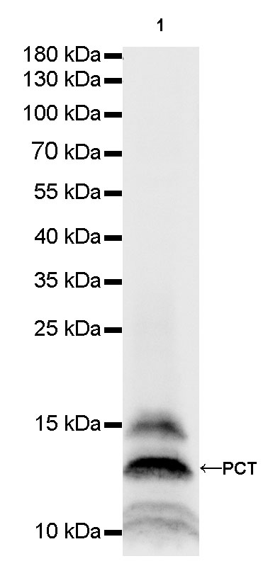
WB result of PCT Rabbit mAb
Primary antibody: PCT Rabbit mAb at 1/1000 dilution
Lane 1: TT whole cell lysate 20 µg
Secondary antibody: Goat Anti-Rabbit IgG, (H+L), HRP conjugated at 1/10000 dilution
Predicted MW: 15 kDa
Observed MW: 13 kDa

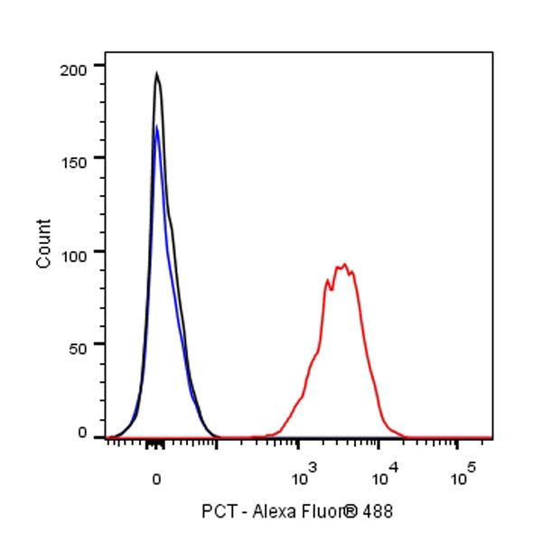
Flow cytometric analysis of 4% PFA fixed 90% methanol permeabilized TT (Human thyroid carcinoma epithelial cell) cells labelling PCT antibody at 1/50 (1 μg) dilution / (red) compared with a Rabbit monoclonal IgG (Black) isotype control and an unlabelled control (cells without incubation with primary antibody and secondary antibody) (Blue). Goat Anti - Rabbit IgG Alexa Fluor® 488 was used as the secondary antibody.
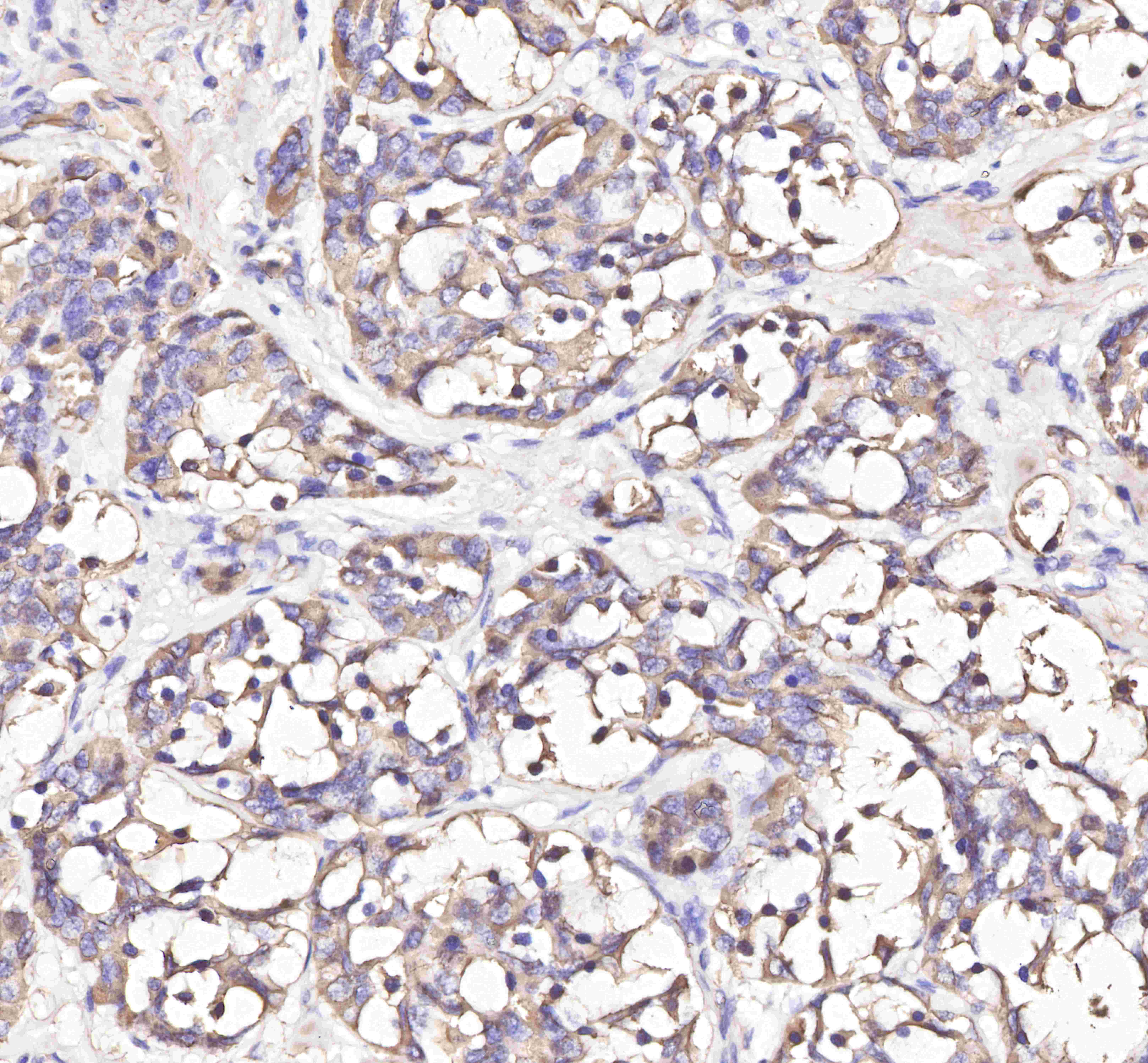
IHC shows positive staining in paraffin-embedded human thyroid medullary carcinoma.
Anti-PCT antibody was used at 1/2000 dilution, followed by a Goat Anti-Rabbit IgG H&L (HRP) ready to use.
Counterstained with hematoxylin.
Heat mediated antigen retrieval with Tris/EDTA buffer pH9.0 was performed before commencing with IHC staining protocol.
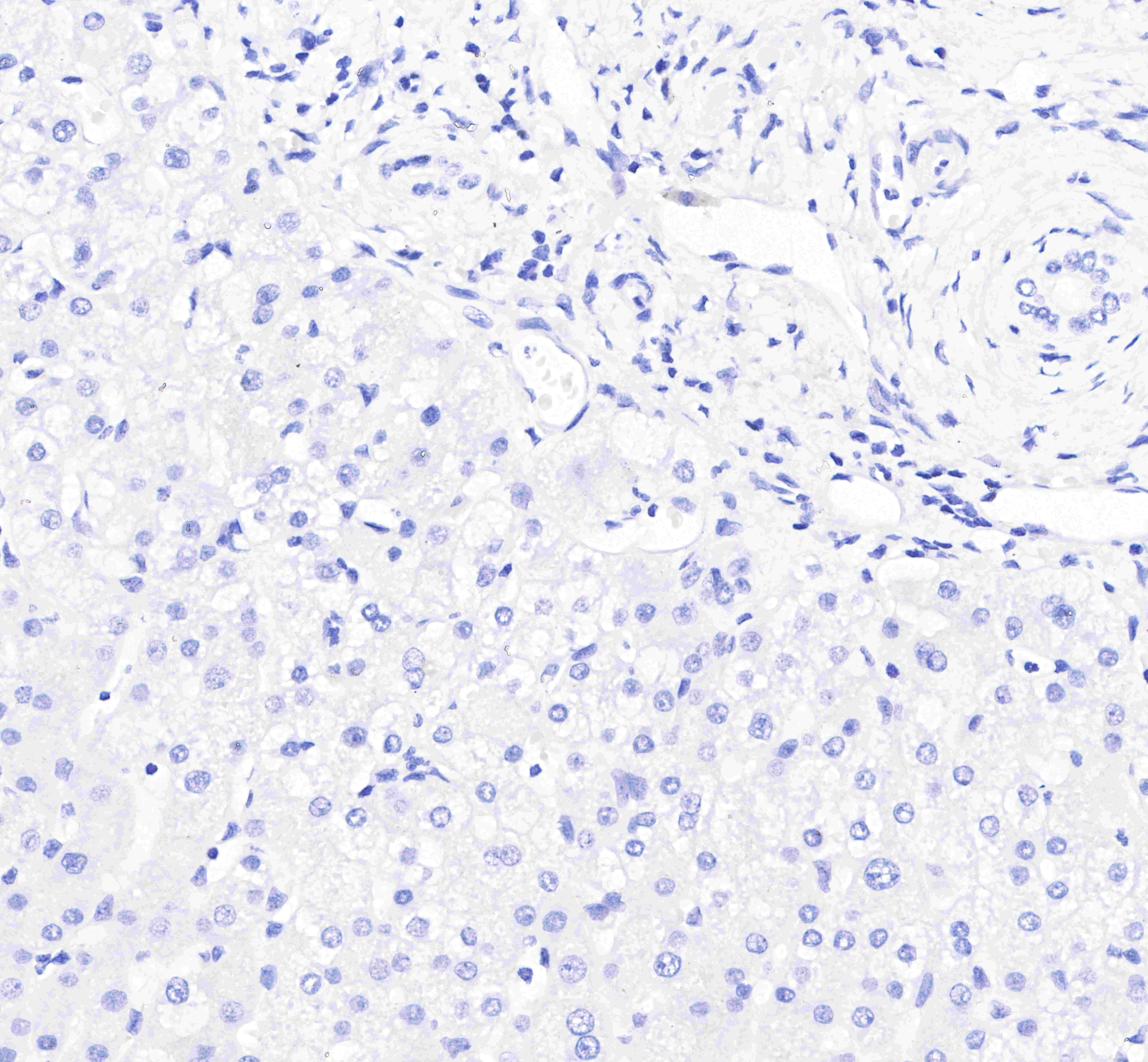
Negative control: IHC shows negative staining in paraffin-embedded human liver.
Anti-PCT antibody was used at 1/2000 dilution, followed by a Goat Anti-Rabbit IgG H&L (HRP) ready to use.
Counterstained with hematoxylin.
Heat mediated antigen retrieval with Tris/EDTA buffer pH9.0 was performed before commencing with IHC staining protocol.
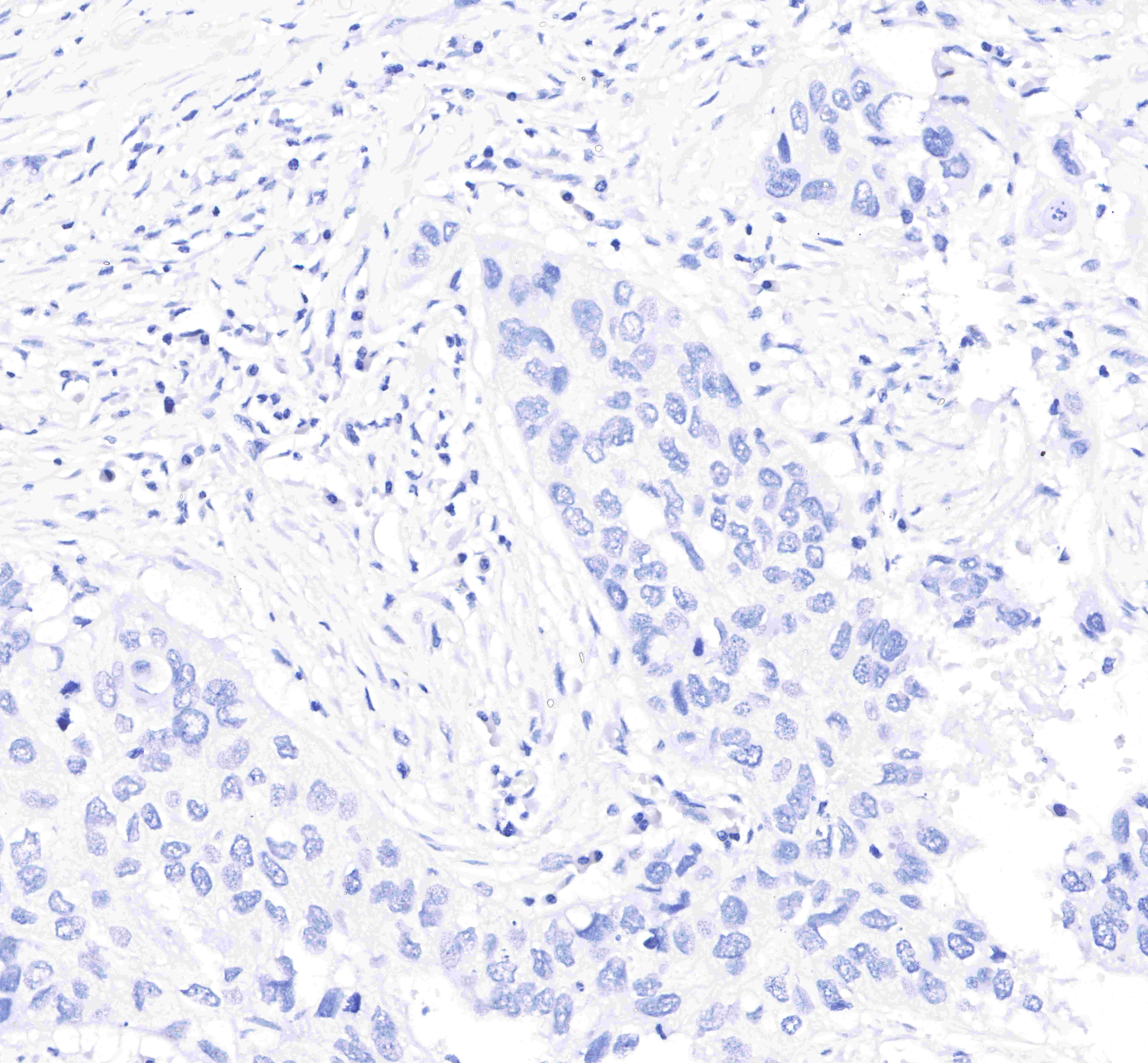
Negative control: IHC shows negative staining in paraffin-embedded human lung squamous cell cancer.
Anti-PCT antibody was used at 1/2000 dilution, followed by a Goat Anti-Rabbit IgG H&L (HRP) ready to use.
Counterstained with hematoxylin.
Heat mediated antigen retrieval with Tris/EDTA buffer pH9.0 was performed before commencing with IHC staining protocol.
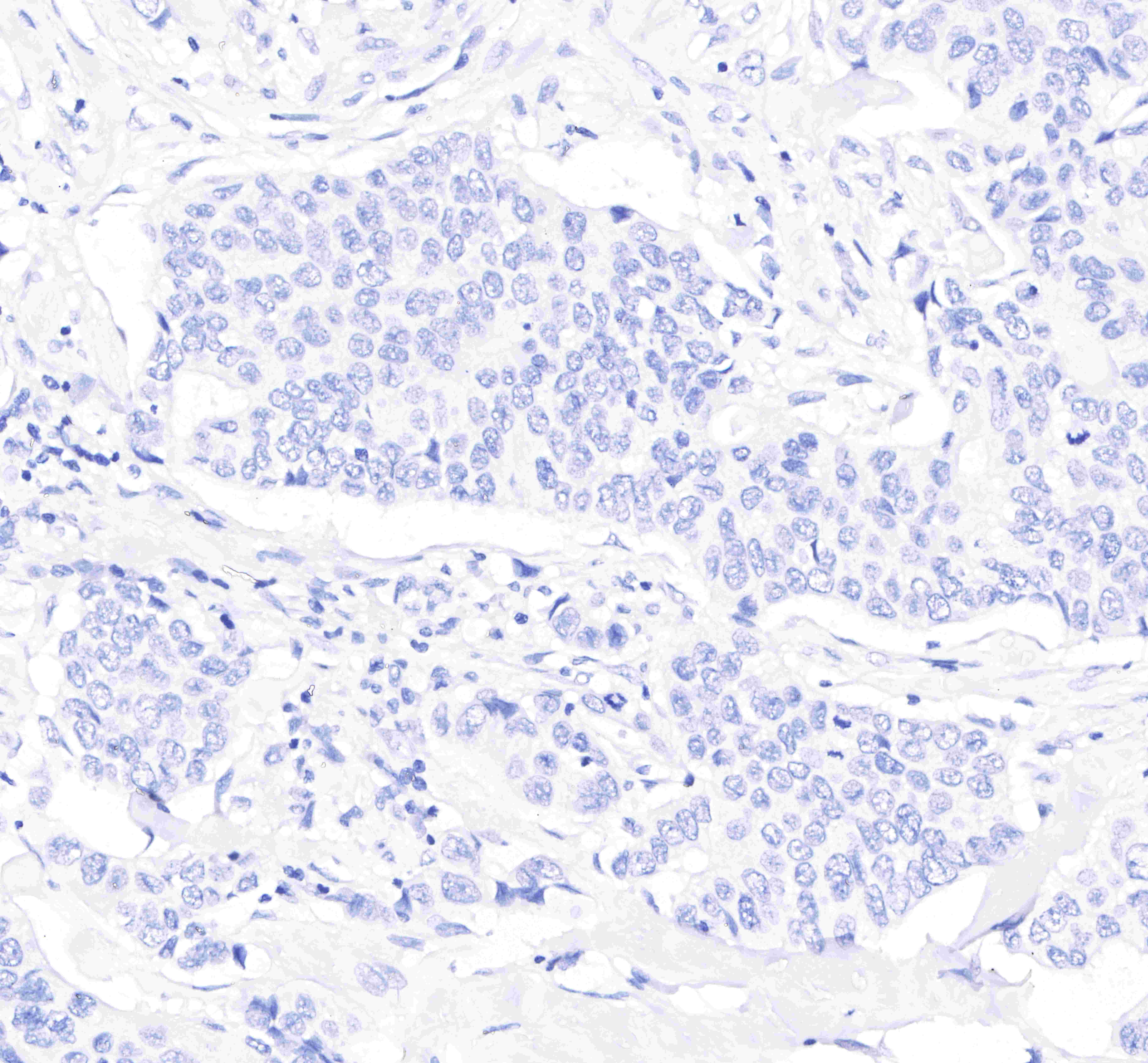
Negative control: IHC shows negative staining in paraffin-embedded human breast cancer.
Anti-PCT antibody was used at 1/2000 dilution, followed by a Goat Anti-Rabbit IgG H&L (HRP) ready to use.
Counterstained with hematoxylin.
Heat mediated antigen retrieval with Tris/EDTA buffer pH9.0 was performed before commencing with IHC staining protocol.