WB result of p16 Rabbit mAb
Primary antibody: p16 Rabbit mAb at 1/1000 dilution
Lane 1: MCF7 whole cell lysate 20 µg
Lane 2: HeLa whole cell lysate 20 µg
Lane 3: HEK-293 whole cell lysate 20 µg
Negative control: MCF7 whole cell lysate
Secondary antibody: Goat Anti-Rabbit IgG, (H+L), HRP conjugated at 1/10000 dilution
Predicted MW: 16 kDa
Observed MW: 16 kDa
(This blot was developed with high sensitivity substrate)
p16 Recombinant Rabbit mAb (S-303-1)
p16 Recombinant Rabbit mAb (S-303-1)
Price:
Regular price
$100 USD
Regular price
Sale price
$100 USD
Unit price
per
For shipping services or bulk orders, you may request a quotation.
Secure checkout with

View full details
Product Details
Product Details
Product Specification
| Host | Rabbit |
| Antigen | p16 |
| Synonyms | Cyclin-dependent kinase inhibitor 2A, Cyclin-dependent kinase 4 inhibitor A (CDK4I), Multiple tumor suppressor 1 (MTS-1), p16-INK4a (p16-INK4; p16INK4A), CDKN2A, CDKN2, MTS1 |
| Immunogen | Recombinant Protein |
| Location | Cytoplasm, Nucleus |
| Accession | P42771 |
| Clone Number | S-303-1 |
| Antibody Type | Recombinant mAb |
| Application | WB, ICC, ICFCM, IP |
| Reactivity | Hu |
| Purification | Protein A |
| Concentration | 0.5 mg/ml |
| Conjugation | Unconjugated |
| Physical Appearance | Liquid |
| Storage Buffer | PBS, 40% Glycerol, 0.05%BSA, 0.03% Proclin 300 |
| Stability & Storage | 12 months from date of receipt / reconstitution, -20 °C as supplied |
Dilution
| application | dilution | species |
| WB | 1:1000 | null |
| IP | 1:50 | null |
| ICC | 1:100 | null |
| ICFCM | 1:500 | null |
Background
p16 (also known as p16INK4a, cyclin-dependent kinase inhibitor 2A, CDKN2A, multiple tumor suppressor 1 and numerous other synonyms), is a protein that slows cell division by slowing the progression of the cell cycle from the G1 phase to the S phase, thereby acting as a tumor suppressor. p16 can be used as a biomarker to improve the histological diagnostic accuracy of grade 3 cervical intraepithelial neoplasia (CIN). p16 is also implicated in the prevention of melanoma, oropharyngeal squamous cell carcinoma, cervical cancer, vulvar cancer and esophageal cancer.
Picture
Picture
Western Blot
FC
Flow cytometric analysis of 4% PFA fixed 90% methanol permeabilized HeLa (Human cervix adenocarcinoma epithelial cell) cells labelling p16 antibody at 1/500 dilution (0.1 μg) / (red) compared with a Rabbit monoclonal IgG (Black) isotype control and an unlabelled control (cells without incubation with primary antibody and secondary antibody) (Blue). Goat Anti - Rabbit IgG Alexa Fluor® 488 was used as the secondary antibody.
IP
p16 Rabbit mAb at 1/50 dilution (1 µg) immunoprecipitating p16 in 0.4 mg HeLa whole cell lysate.
Western blot was performed on the immunoprecipitate using p16 Rabbit mAb at 1/1000 dilution.
Secondary antibody (HRP) for IP was used at 1/400 dilution.
Lane 1: HeLa whole cell lysate 20 µg (Input)
Lane 2: p16 Rabbit mAb IP in HeLa whole cell lysate
Lane 3: Rabbit monoclonal IgG IP in HeLa whole cell lysate
Predicted MW: 16 kDa
Observed MW: 16 kDa
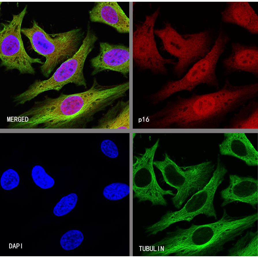
Immunocytochemistry
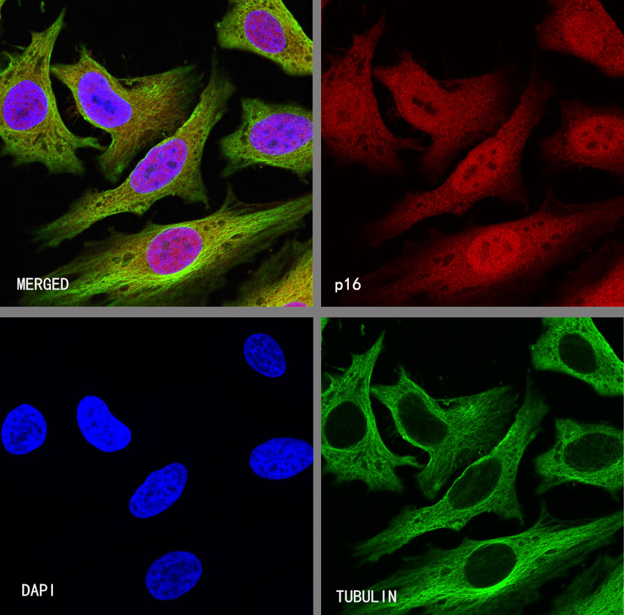
ICC shows positive staining in HeLa cells. Anti-p16 antibody was used at 1/100 dilution (Red) and incubated overnight at 4°C. Goat polyclonal Antibody to Rabbit IgG - H&L (Alexa Fluor® 594) was used as secondary antibody at 1/1000 dilution. The cells were fixed with 4% PFA and permeabilized with 0.1% PBS-Triton X-100. Nuclei were counterstained with DAPI (Blue).Counterstain with tubulin (Green).