WB result of p14ARF Rabbit mAb
Primary antibody: p14ARF Rabbit mAb at 1/1000 dilution
Lane 1: SK-OV-3 whole cell lysate 20 µg
Lane 2: HeLa whole cell lysate 20 µg
Lane 3: Saos-2 whole cell lysate 20 µg
Negative control: SK-OV-3 whole cell lysate
Secondary antibody: Goat Anti-Rabbit IgG, (H+L), HRP conjugated at 1/10000 dilution
Predicted MW: 14 kDa
Observed MW: 14 kDa
(This blot was developed with high sensitivity substrate)
p14ARF Recombinant Rabbit mAb (S-R244)
p14ARF Recombinant Rabbit mAb (S-R244)
Price:
Regular price
$100 USD
Regular price
Sale price
$100 USD
Unit price
per
For shipping services or bulk orders, you may request a quotation.
Secure checkout with

View full details
Product Details
Product Details
Product Specification
| Host | Rabbit |
| Antigen | p14ARF |
| Synonyms | Tumor suppressor ARF, Alternative reading frame (ARF), Cyclin-dependent kinase inhibitor 2A, CDKN2A, MLM |
| Location | Nucleus |
| Accession | Q8N726 |
| Clone Number | S-R244 |
| Antibody Type | Recombinant mAb |
| Application | WB, IHC-P, ICC, ICFCM, IP |
| Reactivity | Hu |
| Purification | Protein A |
| Concentration | 0.5 mg/ml |
| Conjugation | Unconjugated |
| Physical Appearance | Liquid |
| Storage Buffer | PBS, 40% Glycerol, 0.05%BSA, 0.03% Proclin 300 |
| Stability & Storage | 12 months from date of receipt / reconstitution, -20 °C as supplied |
Dilution
| application | dilution | species |
| WB | 1:1000 | null |
| IHC | 1:100 | null |
| ICFCM | 1:500 | null |
| ICC | 1:500 | null |
| IP | 1:50 | null |
Background
p14ARF (also called ARF tumor suppressor, ARF, p14ARF) is an alternate reading frame protein product of the CDKN2A locus (i.e. INK4a/ARF locus). p14ARF is induced in response to elevated mitogenic stimulation, such as aberrant growth signaling from MYC and Ras (protein). It accumulates mainly in the nucleolus where it forms stable complexes with NPM or Mdm2. These interactions allow p14ARF to act as a tumor suppressor by inhibiting ribosome biogenesis or initiating p53-dependent cell cycle arrest and apoptosis, respectively.
Picture
Picture
Western Blot
FC
Flow cytometric analysis of 4% PFA fixed 90% methanol permeabilized SK-OV-3 (Human ovarian cancer epithelial cell, left) / HeLa (Human cervix adenocarcinoma epithelial cell, right) cells labelling p14ARF antibody at 1/500 dilution (0.1 μg) / (red) compared with a Rabbit monoclonal IgG (Black) isotype control and an unlabelled control (cells without incubation with primary antibody and secondary antibody) (Blue). Goat Anti - Rabbit IgG Alexa Fluor® 488 was used as the secondary antibody.
Negative control: SK-OV-3
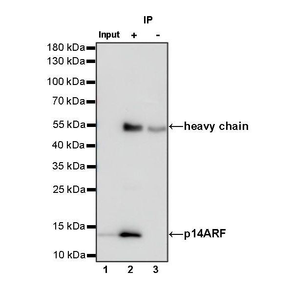
IP
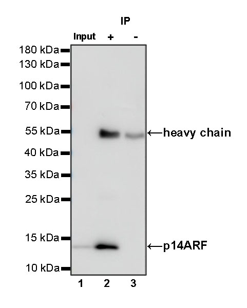
p14ARF Rabbit mAb at 1/50 dilution (1 µg) immunoprecipitating p14ARF in 0.4 mg HeLa whole cell lysate.
Western blot was performed on the immunoprecipitate using p14ARF Rabbit mAb at 1/1000 dilution.
Secondary antibody (HRP) for IP was used at 1/400 dilution.
Lane 1: HeLa whole cell lysate 20 µg (Input)
Lane 2: p14ARF Rabbit mAb IP in HeLa whole cell lysate
Lane 3: Rabbit monoclonal IgG IP in HeLa whole cell lysate
Predicted MW: 14 kDa
Observed MW: 14 kDa
Immunohistochemistry
IHC shows positive staining in paraffin-embedded human cervical squamous cell carcinoma. Anti-p14ARF antibody was used at 1/100 dilution, followed by a HRP Polymer for Mouse & Rabbit IgG (ready to use). Counterstained with hematoxylin. Heat mediated antigen retrieval with Tris/EDTA buffer pH9.0 was performed before commencing with IHC staining protocol.
IHC shows positive staining in paraffin-embedded human lung adenocarcinoma. Anti-p14ARF antibody was used at 1/100 dilution, followed by a HRP Polymer for Mouse & Rabbit IgG (ready to use). Counterstained with hematoxylin. Heat mediated antigen retrieval with Tris/EDTA buffer pH9.0 was performed before commencing with IHC staining protocol.
IHC shows positive staining in paraffin-embedded human ovarian carcinoma. Anti-p14ARF antibody was used at 1/100 dilution, followed by a HRP Polymer for Mouse & Rabbit IgG (ready to use). Counterstained with hematoxylin. Heat mediated antigen retrieval with Tris/EDTA buffer pH9.0 was performed before commencing with IHC staining protocol.
Negative control: IHC shows negative staining in paraffin-embedded human skeletal muscle. Anti-p14ARF antibody was used at 1/100 dilution, followed by a HRP Polymer for Mouse & Rabbit IgG (ready to use). Counterstained with hematoxylin. Heat mediated antigen retrieval with Tris/EDTA buffer pH9.0 was performed before commencing with IHC staining protocol.
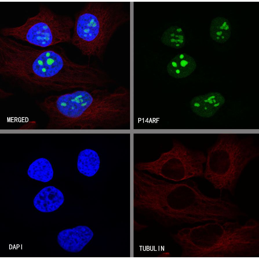
Immunocytochemistry
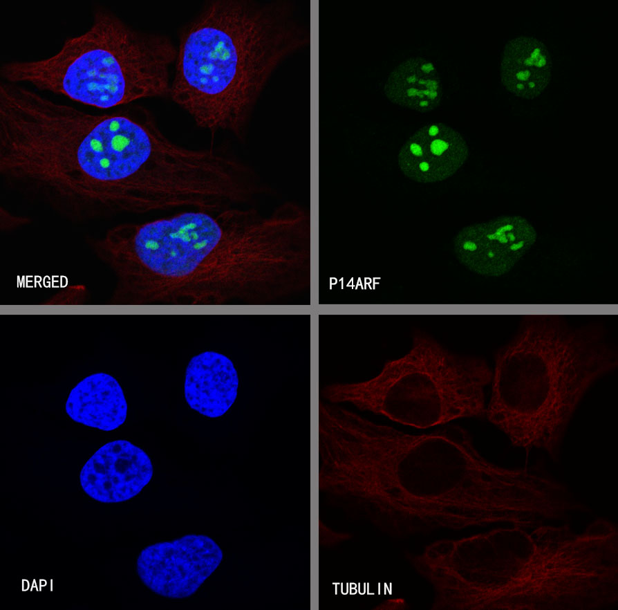
ICC shows positive staining in HeLa cells. Anti-p14ARF antibody was used at 1/500 dilution (Green) and incubated overnight at 4°C. Goat polyclonal Antibody to rabbit IgG - H&L (Alexa Fluor® 488) was used as secondary antibody at 1/1000 dilution. The cells were fixed with 4% PFA and permeabilized with 0.1% PBS-Triton X-100. Nuclei were counterstained with DAPI (Blue).Counterstain with tubulin (Red).
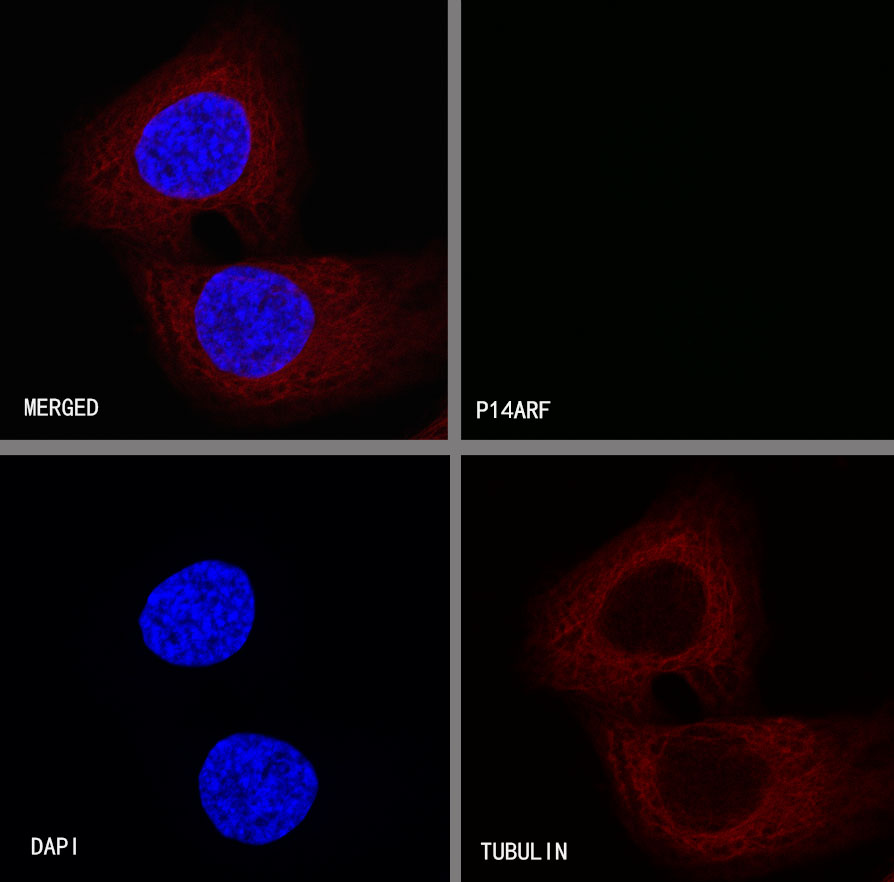
Negative control:ICC shows negative staining in SK-OV-3 cells. Anti-p14ARF antibody was used at 1/500 dilution and incubated overnight at 4°C. Goat polyclonal Antibody to rabbit IgG - H&L (Alexa Fluor® 488) was used as secondary antibody at 1/1000 dilution. The cells were fixed with 4% PFA and permeabilized with 0.1% PBS-Triton X-100. Nuclei were counterstained with DAPI (Blue).Counterstain with tubulin (Red).