WB result of HP1α Recombinant Rabbit mAb
Primary antibody: HP1α Recombinant Rabbit mAb at 1/1000 dilution
Lane 1: HeLa whole cell lysate 20 µg
Lane 2: 293T whole cell lysate 20 µg
Lane 3: MCF7 whole cell lysate 20 µg
Secondary antibody: Goat Anti-Rabbit IgG, (H+L), HRP conjugated at 1/10000 dilution Predicted MW: 22 kDa
Observed MW: 22 kDa
HP1α Recombinant Rabbit mAb (S-1462-53)
HP1α Recombinant Rabbit mAb (S-1462-53)
Price:
Regular price
$100 USD
Regular price
Sale price
$100 USD
Unit price
per
For shipping services or bulk orders, you may request a quotation.
Secure checkout with

View full details
Product Details
Product Details
Product Specification
| Host | Rabbit |
| Antigen | HP1α |
| Synonyms | Chromobox protein homolog 5; Antigen p25; Heterochromatin protein 1 homolog alpha (HP1 alpha); CBX5; HP1A |
| Immunogen | Synthetic Peptide |
| Location | Nucleus |
| Accession | P45973 |
| Clone Number | S-1462-53 |
| Antibody Type | Recombinant mAb |
| Isotype | IgG |
| Application | WB, ICC, ICFCM, ChIP |
| Reactivity | Hu, Ms, Rt |
| Positive Sample | HeLa, 293T, MCF7, NIH/3T3, C6 |
| Purification | Protein A |
| Concentration | 0.5 mg/ml |
| Conjugation | Unconjugated |
| Physical Appearance | Liquid |
| Storage Buffer | PBS, 40% Glycerol, 0.05% BSA, 0.03% Proclin 300 |
| Stability & Storage | 12 months from date of receipt / reconstitution, -20 °C as supplied |
Dilution
| application | dilution | species |
| WB | 1:1000 | Hu, Rt |
| ICC | 1:500 | Hu, Ms |
| ChIP | 1:20-1:50 |
Background
HP1α (heterochromatin protein 1 alpha) is a structural chromosomal protein that plays a multifaceted role in chromatin regulation and gene expression. It is primarily associated with heterochromatin, a condensed form of chromatin that promotes gene silencing, particularly at telomeres and centromeres. HP1α binds to the histone modification H3K9me3 (histone H3 lysine 9 trimethylation) via its N-terminal chromodomain, which is crucial for the assembly and maintenance of heterochromatin. This binding helps to compact chromatin, control gene expression, and maintain genome integrity. In addition to its role in gene silencing, HP1α also has the capacity to promote gene expression. It can impact the expression of genes in euchromatin as well as heterochromatic genes. The mechanisms by which HP1α can positively regulate gene expression include maintenance of heterochromatin structure and facilitating transcriptional elongation. HP1α is also involved in the induction of heat-shock genes and has been shown to interact directly with RNA polymerase II and heterogeneous nuclear ribonuclear proteins (hnRNPs), suggesting a role in RNA processing.
Picture
Picture
Western Blot
WB result of HP1α Recombinant Rabbit mAb
Primary antibody: HP1α Recombinant Rabbit mAb at 1/1000 dilution
Lane 1: C6 whole cell lysate 20 µg
Secondary antibody: Goat Anti-Rabbit IgG, (H+L), HRP conjugated at 1/10000 dilution Predicted MW: 22 kDa
Observed MW: 22 kDa
FC
Flow cytometric analysis of 4% PFA fixed 90% methanol permeabilized HeLa (Human cervix adenocarcinoma epithelial cell) labelling HP1 alpha antibody at 1/500 dilution (0.1 μg)/ (Red) compared with a Rabbit monoclonal IgG (Black) isotype control and an unlabelled control (cells without incubation with primary antibody and secondary antibody) (Blue). Goat Anti - Rabbit IgG Alexa Fluor® 488 was used as the secondary antibody.
Immunocytochemistry
ICC shows positive staining in HeLa cells. Anti- HP1 alpha antibody was used at 1/500 dilution (Green) and incubated overnight at 4°C. Goat polyclonal Antibody to Rabbit IgG - H&L (Alexa Fluor® 488) was used as secondary antibody at 1/1000 dilution. The cells were fixed with 4% PFA and permeabilized with 0.1% PBS-Triton X-100. Nuclei were counterstained with DAPI (Blue). Counterstain with tubulin (Red).
ICC shows positive staining in NIH/3T3 cells. Anti- HP1 alpha antibody was used at 1/500 dilution (Green) and incubated overnight at 4°C. Goat polyclonal Antibody to Rabbit IgG - H&L (Alexa Fluor® 488) was used as secondary antibody at 1/1000 dilution. The cells were fixed with 4% PFA and permeabilized with 0.1% PBS-Triton X-100. Nuclei were counterstained with DAPI (Blue). Counterstain with tubulin (Red).
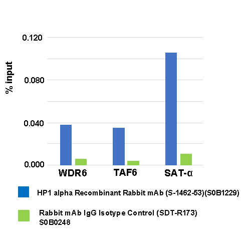
ChIP
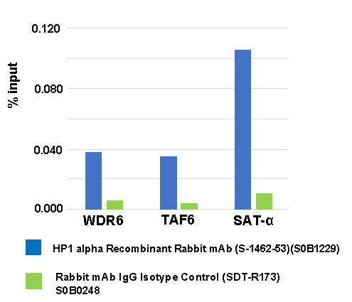
Chromatin immunoprecipitation (ChIP) was performed on HeLa cells cross - linked with 1%
formaldehyde for 10 min, then chromatin was fragmented by sonication. Parallel reactions used HP1 alpha Recombinant Rabbit mAb (S-1462-53)and IgG Isotype Control (SDT-R173) at 1:50 for immunoprecipitation.
Post - immunoprecipitation, both samples were washed, eluted, and cross - links reversed. Purified DNA was analyzed by qPCR.
qPCR (%input: immunoprecipitated DNA/input DNA) showed the enrichment of WDR6, TAF6 and SAT-α in
HP1 alpha Recombinant Rabbit mAb (S-1462-53)-immunoprecipitated sample.