
WB result of HexokinaseⅠRecombinant Rabbit mAb
Primary antibody: HexokinaseⅠRecombinant Rabbit mAb at 1/1000 dilution
Lane 1: 293T whole cell lysate 20 µg
Lane 2: HeLa whole cell lysate 20 µg
Lane 3: Jurkat whole cell lysate 20 µg
Lane 4: MCF7 whole cell lysate 20 µg
Lane 5: U-87 MG whole cell lysate 20 µg
Secondary antibody: Goat Anti-Rabbit IgG, (H+L), HRP conjugated at 1/10000 dilution Predicted MW: 102 kDa
Observed MW: 110 kDa
HexokinaseⅠRecombinant Rabbit mAb (S-868-113)
HexokinaseⅠRecombinant Rabbit mAb (S-868-113)
Price:
Regular price
$100 USD
Regular price
Sale price
$100 USD
Unit price
per
For shipping services or bulk orders, you may request a quotation.
Secure checkout with

View full details
Product Details
Product Details
Product Specification
| Host | Rabbit |
| Antigen | HexokinaseⅠ |
| Synonyms | Brain form hexokinase; Hexokinase type I (HK I); Hexokinase-A; HK1 |
| Immunogen | Synthetic Peptide |
| Location | Mitochondrion outer membrane |
| Accession | P19367 |
| Clone Number | S-868-113 |
| Antibody Type | Recombinant mAb |
| Isotype | IgG |
| Application | WB, ICC, ICFCM |
| Reactivity | Hu, Ms |
| Positive Sample | 293T, HeLa, Jurkat, MCF7, U-87 MG, NIH/3T3, C2C12 |
| Purification | Protein A |
| Concentration | 0.5 mg/ml |
| Conjugation | Unconjugated |
| Physical Appearance | Liquid |
| Storage Buffer | PBS, 40% Glycerol, 0.05% BSA, 0.03% Proclin 300 |
| Stability & Storage | 12 months from date of receipt / reconstitution, -20 °C as supplied |
Dilution
| application | dilution | species |
| WB | 1:1000 | Hu, Ms |
| ICC | 1:500 | Hu |
| ICFCM | 1:50 |
Background
Hexokinase I (HK-I) is a subtype of the hexokinase family. The primary function of HK-I is to regulate the entry of glucose into the glycolytic pathway and generate ATP. It is firmly bound to mitochondria and, in the brain, HK-I catalyzes the phosphorylation of glucose, determining the rate of glycolysis in the brain. HK-I also acts as a coordinating enzyme to prevent an imbalance between glycolysis and mitochondrial oxidative reactions, which could lead to the accumulation of lactic acid. Additionally, in hematopoietic cell lines, HK-I is co-expressed with GLUT1, increasing the level of glucose phosphorylation and the level of cellular nicotinamide adenine dinucleotide phosphate (NADPH), thereby exerting an anti-apoptotic effect.
Picture
Picture
Western Blot
WB result of HexokinaseⅠRecombinant Rabbit mAb
Primary antibody: HexokinaseⅠRecombinant Rabbit mAb at 1/1000 dilution
Lane 1: NIH/3T3 whole cell lysate 20 µg
Lane 2: C2C12 whole cell lysate 20 µg
Secondary antibody: Goat Anti-Rabbit IgG, (H+L), HRP conjugated at 1/10000 dilution Predicted MW: 102 kDa
Observed MW: 110 kDa
FC
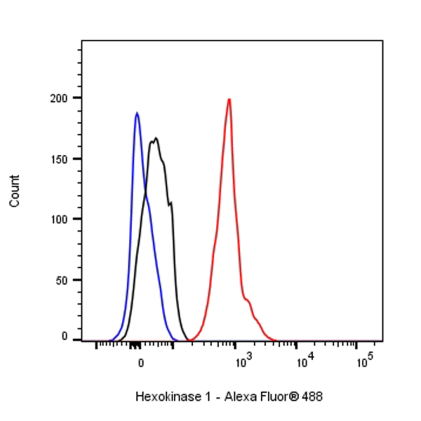
Flow cytometric analysis of 4% PFA fixed 90% methanol permeabilized HeLa (Human cervix adenocarcinoma epithelial cell) cells labelling Hexokinase 1 antibody at 1/50 dilution (1 μg)/ (Red) compared with a Rabbit monoclonal IgG (Black) isotype control and an unlabelled control (cells without incubation with primary antibody and secondary antibody) (Blue). Goat Anti - Rabbit IgG Alexa Fluor® 488 was used as the secondary antibody.
Immunocytochemistry
ICC shows positive staining in HeLa cells. Anti- Hexokinase 1 antibody was used at 1/500 dilution (Green) and incubated overnight at 4°C. Goat polyclonal Antibody to Rabbit IgG - H&L (Alexa Fluor® 488) was used as secondary antibody at 1/1000 dilution. The cells were fixed with 100% ice-cold methanol and permeabilized with 0.1% PBS-Triton X-100. Nuclei were counterstained with DAPI (Blue). Counterstain with tubulin (Red).
