WB result of H-Ras Recombinant Rabbit mAb
Primary antibody: H-Ras Recombinant Rabbit mAb at 1/1000 dilution
Lane 1: HeLa whole cell lysate 20 µg
Lane 2: MCF-7 whole cell lysate 20 µg
Lane 3: Jurkat whole cell lysate 20 µg
Lane 4: K562 whole cell lysate 20 µg
Lane 5: A431 whole cell lysate 20 µg
Secondary antibody: Goat Anti-rabbit IgG, (H+L), HRP conjugated at 1/10000 dilution
Predicted MW: 21 kDa
Observed MW: 21 kDa
H-Ras Recombinant Rabbit mAb (S-1017-79)
H-Ras Recombinant Rabbit mAb (S-1017-79)
Price:
Regular price
$100 USD
Regular price
Sale price
$100 USD
Unit price
per
For shipping services or bulk orders, you may request a quotation.
Secure checkout with

View full details
Product Details
Product Details
Product Specification
| Host | Rabbit |
| Antigen | H-Ras |
| Synonyms | GTPase HRas, H-Ras-1, Ha-Ras, Transforming protein p21, c-H-ras, p21ras |
| Immunogen | Synthetic Peptide |
| Location | Cell membrane |
| Accession | P01112 |
| Clone Number | S-1017-79 |
| Antibody Type | Recombinant mAb |
| Isotype | IgG |
| Application | WB, ICC, ICFCM, IP |
| Reactivity | Hu, Ms, Rt |
| Predicted Reactivity | Ys, Or, Xe, Dr, GP, Av, Fs, Zf, Tk, C.el, Hm |
| Purification | Protein A |
| Concentration | 0.5 mg/ml |
| Conjugation | Unconjugated |
| Physical Appearance | Liquid |
| Storage Buffer | PBS, 40% Glycerol, 0.05% BSA, 0.03% Proclin 300 |
| Stability & Storage | 12 months from date of receipt / reconstitution, -20 °C as supplied |
Dilution
| application | dilution | species |
| WB | 1:1000 | |
| ICC | 1:100 | |
| ICFCM | 1:50 | |
| IP | 1:50 |
Background
GTPase HRas, from "Harvey Rat sarcoma virus", also known as transforming protein p21 is an enzyme that in humans is encoded by the HRAS gene. The HRAS protein is a GTPase and is an early player in many signal transduction pathways and is usually associated with cell membranes due to the presence of an isoprenyl group on its C-terminus. HRAS acts as a molecular on/off switch, once it is turned on it recruits and activates proteins necessary for the propagation of the receptor's signal, such as c-Raf and PI 3-kinase. HRAS binds to GTP in the active state and possesses an intrinsic enzymatic activity that cleaves the terminal phosphate of this nucleotide converting it to GDP. Upon conversion of GTP to GDP, HRAS is turned off. The rate of conversion is usually slow but can be sped up dramatically by an accessory protein of the GTPase activating protein (GAP) class, for example RasGAP. HRAS has been shown to be a proto-oncogene.
Picture
Picture
Western Blot
WB result of H-Ras Recombinant Rabbit mAb
Primary antibody: H-Ras Recombinant Rabbit mAb at 1/1000 dilution
Lane 1: RAW264.7 whole cell lysate 20 µg
Secondary antibody: Goat Anti-rabbit IgG, (H+L), HRP conjugated at 1/10000 dilution
Predicted MW: 21 kDa
Observed MW: 21 kDa
WB result of H-Ras Recombinant Rabbit mAb
Primary antibody: H-Ras Recombinant Rabbit mAb at 1/1000 dilution
Lane 1: NIH/3T3 whole cell lysate 20 µg
Secondary antibody: Goat Anti-rabbit IgG, (H+L), HRP conjugated at 1/10000 dilution
Predicted MW: 21 kDa
Observed MW: 21 kDa
FC
Flow cytometric analysis of 4% PFA fixed 90% methanol permeabilized Jurkat (Human T cell leukemia T lymphocyte) labelling H-Ras antibody at 1/50 dilution (1 μg)/ (Red) compared with a Rabbit monoclonal IgG (Black) isotype control and an unlabelled control (cells without incubation with primary antibody and secondary antibody) (Blue). Goat Anti - Rabbit IgG Alexa Fluor® 488 was used as the secondary antibody.
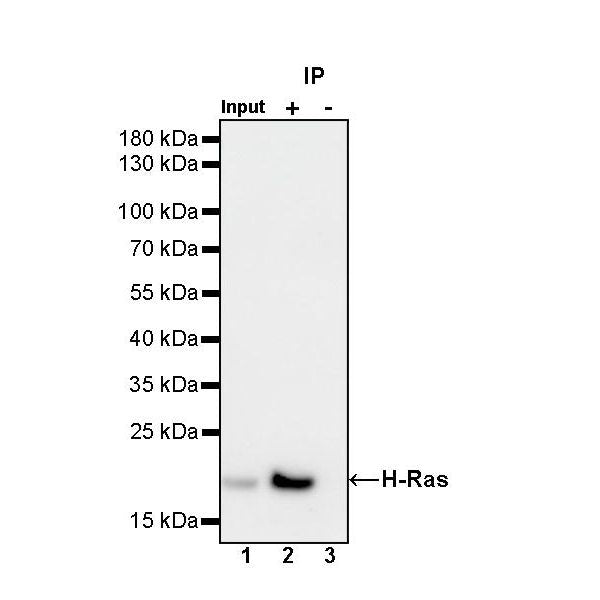
IP
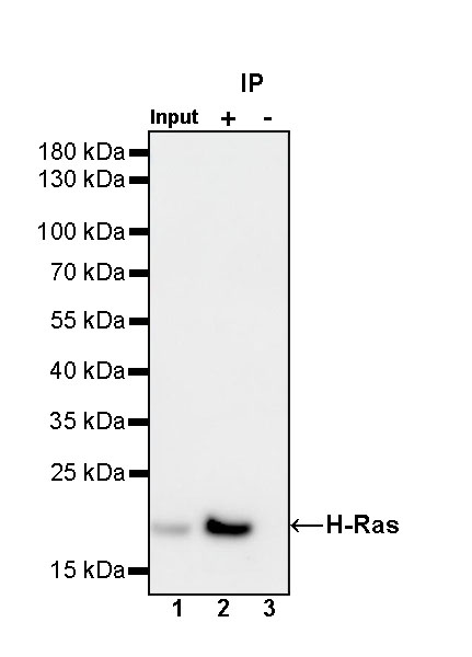
H-Ras Rabbit mAb at 1/50 dilution (1 µg) immunoprecipitating H-Ras in 0.4 mg Jurkat whole cell lysate.
Western blot was performed on the immunoprecipitate using H-Ras Rabbit mAb at 1/1000 dilution.
Secondary antibody (HRP) for IP was used at 1/1000 dilution.
Lane 1: Jurkat whole cell lysate 20 µg (Input)
Lane 2: H-Ras Rabbit mAb IP in Jurkat whole cell lysate
Lane 3: Rabbit monoclonal IgG IP in Jurkat whole cell lysate
Predicted MW: 21 kDa
Observed MW: 21 kDa
Immunocytochemistry
ICC shows positive staining in Jurkat cells. Anti- H-Ras antibody was used at 1/100 dilution (Green) and incubated overnight at 4°C. Goat polyclonal Antibody to Rabbit IgG - H&L (Alexa Fluor® 488) was used as secondary antibody at 1/1000 dilution. The cells were fixed with 4% PFA and permeabilized with 0.1% PBS-Triton X-100. Nuclei were counterstained with DAPI (Blue). Counterstain with tubulin (Red).