WB result of FAK Recombinant Rabbit mAb
Primary antibody: FAK Recombinant Rabbit mAb at 1/1000 dilution
Lane 1: HeLa whole cell lysate 20 µg
Lane 2: HEK-293 whole cell lysate 20 µg
Lane 3: K562 whole cell lysate 20 µg
Lane 4: MCF-7 whole cell lysate 20 µg
Secondary antibody: Goat Anti-rabbit IgG, (H+L), HRP conjugated at 1/10000 dilution
Predicted MW: 119 kDa
Observed MW: 120 kDa
FAK Recombinant Rabbit mAb (S-1180-10)
FAK Recombinant Rabbit mAb (S-1180-10)
Price:
Regular price
$100 USD
Regular price
Sale price
$100 USD
Unit price
per
For shipping services or bulk orders, you may request a quotation.
Secure checkout with

View full details
Product Details
Product Details
Product Specification
| Host | Rabbit |
| Antigen | FAK |
| Synonyms | Focal adhesion kinase 1, FADK 1, Focal adhesion kinase-related nonkinase (FRNK), Protein phosphatase 1 regulatory subunit 71 (PPP1R71), Protein-tyrosine kinase 2, p125FAK, pp125FAK, PTK2, FAK1 |
| Immunogen | Synthetic Peptide |
| Location | Cytoplasm, Nucleus, Cell membrane |
| Accession | Q05397 |
| Clone Number | S-1180-10 |
| Antibody Type | Recombinant mAb |
| Isotype | IgG |
| Application | WB, IHC-P, ICC, FCM, IP |
| Reactivity | Hu, Ms, Rt |
| Predicted Reactivity | Av |
| Purification | Protein A |
| Concentration | 0.5 mg/ml |
| Conjugation | Unconjugated |
| Physical Appearance | Liquid |
| Storage Buffer | PBS, 40% Glycerol, 0.05% BSA, 0.03% Proclin 300 |
| Stability & Storage | 12 months from date of receipt / reconstitution, -20 °C as supplied |
Dilution
| application | dilution | species |
| WB | 1:1000 | |
| IHC-P | 1:200 | |
| ICC | 1:100-1:500 | |
| ICFCM | 1:50 | |
| IP | 1:50 |
Background
FAK protein, also known as Focal Adhesion Kinase (FAK), PTK2 (Protein Tyrosine Kinase 2), or FADK, is a non-receptor protein tyrosine kinase belonging to the PTK superfamily. FAK plays a central role in cellular signal transduction, serving as a hub for integrating signals from integrins, growth factors, mechanical stimuli, and more. It activates intracellular signaling pathways such as PI3K/Akt and Ras/MAPK, regulating cell growth, migration, adhesion, spreading, actin cytoskeleton reorganization, focal adhesion formation and disassembly, cell cycle progression, proliferation, and apoptosis. Additionally, FAK is intimately involved in embryonic development, tumorigenesis, and metastasis.
Picture
Picture
Western Blot
WB result of FAK Recombinant Rabbit mAb
Primary antibody: FAK Recombinant Rabbit mAb at 1/1000 dilution
Lane 1: NIH/3T3 whole cell lysate 20 µg
Lane 2: mouse brain lysate 20 µg
Secondary antibody: Goat Anti-rabbit IgG, (H+L), HRP conjugated at 1/10000 dilution
Predicted MW: 119 kDa
Observed MW: 120 kDa
WB result of FAK Recombinant Rabbit mAb
Primary antibody: FAK Recombinant Rabbit mAb at 1/1000 dilution
Lane 1: C6 whole cell lysate 20 µg
Lane 2: rat brain lysate 20 µg
Secondary antibody: Goat Anti-rabbit IgG, (H+L), HRP conjugated at 1/10000 dilution
Predicted MW: 119 kDa
Observed MW: 120 kDa
FC
Flow cytometric analysis of 4% PFA fixed 90% methanol permeabilized NIH/3T3 (Mouse embryonic fibroblast) labelling FAK antibody at 1/50 dilution (1 μg)/ (Red) compared with a Rabbit monoclonal IgG (Black) isotype control and an unlabelled control (cells without incubation with primary antibody and secondary antibody) (Blue). Goat Anti - Rabbit IgG Alexa Fluor® 488 was used as the secondary antibody.
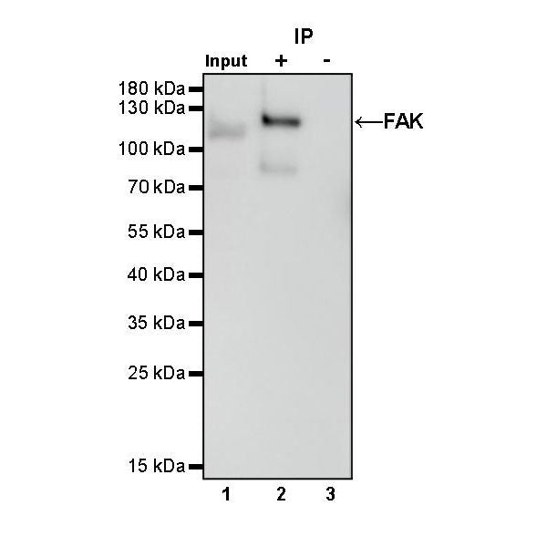
IP

FAK Rabbit mAb at 1/50 dilution (1 µg) immunoprecipitating FAK in 0.4 mg HeLa whole cell lysate.
Western blot was performed on the immunoprecipitate using FAK Rabbit mAb at 1/1000 dilution.
Secondary antibody (HRP) for IP was used at 1/1000 dilution.
Lane 1: HeLa whole cell lysate 20 µg (Input)
Lane 2: FAK Rabbit mAb IP in HeLa whole cell lysate
Lane 3: Rabbit monoclonal IgG IP in HeLa whole cell lysate
Predicted MW: 119 kDa
Observed MW: 120 kDa
Immunohistochemistry
IHC shows positive staining in paraffin-embedded human tonsil. Anti-FAK antibody was used at 1/200 dilution, followed by a HRP Polymer for Mouse & Rabbit IgG (ready to use). Counterstained with hematoxylin. Heat mediated antigen retrieval with Tris/EDTA buffer pH9.0 was performed before commencing with IHC staining protocol.
IHC shows positive staining in paraffin-embedded human lung squamous cell carcinoma. Anti-FAK antibody was used at 1/200 dilution, followed by a HRP Polymer for Mouse & Rabbit IgG (ready to use). Counterstained with hematoxylin. Heat mediated antigen retrieval with Tris/EDTA buffer pH9.0 was performed before commencing with IHC staining protocol.
IHC shows positive staining in paraffin-embedded mouse cerebral cortex. Anti-FAK antibody was used at 1/200 dilution, followed by a HRP Polymer for Mouse & Rabbit IgG (ready to use). Counterstained with hematoxylin. Heat mediated antigen retrieval with Tris/EDTA buffer pH9.0 was performed before commencing with IHC staining protocol.
IHC shows positive staining in paraffin-embedded rat cerebral cortex. Anti-FAK antibody was used at 1/200 dilution, followed by a HRP Polymer for Mouse & Rabbit IgG (ready to use). Counterstained with hematoxylin. Heat mediated antigen retrieval with Tris/EDTA buffer pH9.0 was performed before commencing with IHC staining protocol.
Immunocytochemistry
ICC shows positive staining in MCF-7 cells. Anti-FAK antibody was used at 1/100 dilution (Green) and incubated overnight at 4°C. Goat polyclonal Antibody to Rabbit IgG - H&L (Alexa Fluor® 488) was used as secondary antibody at 1/1000 dilution. The cells were fixed with 4% PFA and permeabilized with 0.1% PBS-Triton X-100. Nuclei were counterstained with DAPI (Blue). Counterstain with tubulin (Red).
ICC shows positive staining in NIH/3T3 cells. Anti-FAK antibody was used at 1/500 dilution (Green) and incubated overnight at 4°C. Goat polyclonal Antibody to Rabbit IgG - H&L (Alexa Fluor® 488) was used as secondary antibody at 1/1000 dilution. The cells were fixed with 100% ice-cold methanol and permeabilized with 0.1% PBS-Triton X-100. Nuclei were counterstained with DAPI (Blue). Counterstain with tubulin (Red).