Flow cytometric analysis of 293T (Human embryonic kidney epithelial cell, left) / Raji (Human Burkitt's lymphoma B lymphocyte, right) cells labelling CD80 antibody at 1/50 dilution (1 μg) / (red) compared with a Rabbit monoclonal IgG (Black) isotype control and an unlabelled control (cells without incubation with primary antibody and secondary antibody) (Blue). Goat Anti-Rabbit IgG Alexa Fluor 488 was used as the secondary antibody. Negative control: 293T
CD80 Recombinant Rabbit mAb (S-288-177)
CD80 Recombinant Rabbit mAb (S-288-177)
Price:
Regular price
$100 USD
Regular price
Sale price
$100 USD
Unit price
per
For shipping services or bulk orders, you may request a quotation.
Secure checkout with

View full details
Product Details
Product Details
Product Specification
| Host | Rabbit |
| Antigen | CD80 |
| Synonyms | T-lymphocyte activation antigen CD80, Activation B7-1 antigen, BB1, CTLA-4 counter-receptor B7.1 (B7) |
| Immunogen | Recombinant Protein |
| Location | Membrane |
| Accession | P33681 |
| Clone Number | S-288-177 |
| Antibody Type | Rabbit mAb |
| Application | ICC, FCM |
| Reactivity | Hu |
| Purification | Protein A |
| Concentration | 0.5 mg/ml |
| Conjugation | Unconjugated |
| Physical Appearance | Liquid |
| Storage Buffer | PBS, 40% Glycerol, 0.05% BSA, 0.03% Proclin 300 |
| Stability & Storage | 12 months from date of receipt / reconstitution, -20 °C as supplied |
Dilution
| application | dilution | species |
| FCM | 1:50 | null |
| ICC | 1:100 | null |
Background
The Cluster of differentiation 80 (also CD80 and B7-1) is a B7, type I membrane protein in the immunoglobulin superfamily, with an extracellular immunoglobulin constant-like domain and a variable-like domain required for receptor binding. It is closely related to CD86, another B7 protein (B7-2), and often works in tandem. Both CD80 and CD86 interact with costimulatory receptors CD28, CTLA-4 (CD152) and the p75 neurotrophin receptor [PMID: 7545666].
Picture
Picture
FC
Immunocytochemistry

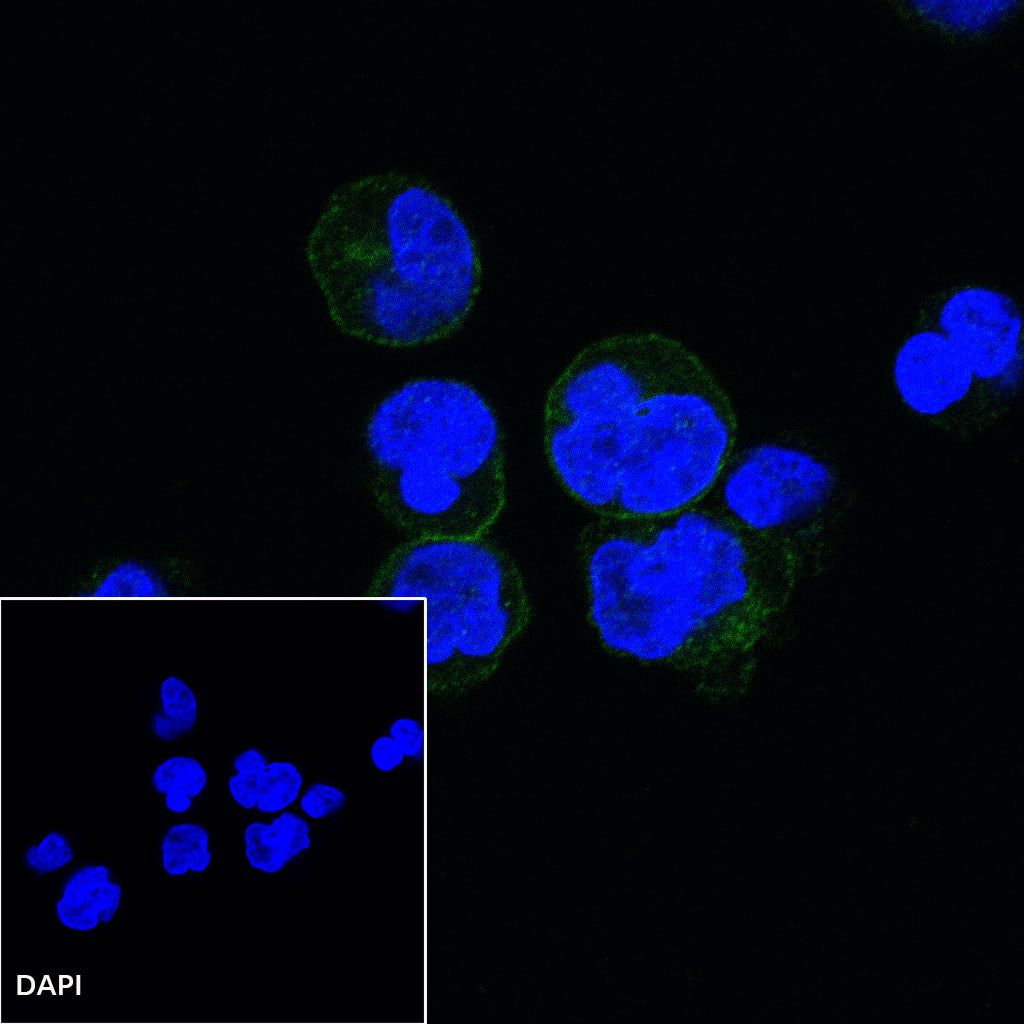
ICC shows positive staining in Raji cells. Anti-CD80 antibody was used at 1/100 dilution (Green) and incubated overnight at 4°C. Goat polyclonal Antibody to Rabbit IgG - H&L (Alexa Fluor® 488) was used as secondary antibody at 1/1000 dilution. The cells were fixed with 100% ice-cold methanol and permeabilized with 0.1% PBS-Triton X-100. Nuclei were counterstained with DAPI (Blue).
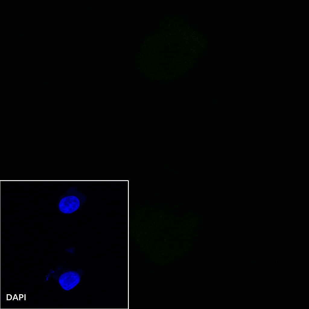
Negative control:ICC shows negative staining in 293T cells. Anti-CD80 antibody was used at 1/100 dilution and incubated overnight at 4°C. Goat polyclonal Antibody to Rabbit IgG - H&L (Alexa Fluor® 488) was used as secondary antibody at 1/1000 dilution. The cells were fixed with 100% ice-cold methanol and permeabilized with 0.1% PBS-Triton X-100. Nuclei were counterstained with DAPI (Blue).