WB result of CD50 Rabbit mAb
Primary antibody: CD50 Rabbit mAb at 1/1000 dilution
Lane 1: 293T whole cell lysate 20 µg
Lane 2: HL-60 whole cell lysate 20 µg
Lane 3: Jurkat whole cell lysate 20 µg
Lane 4: Ramos whole cell lysate 20 µg
Negative control: 293T whole cell lysate
Low expression control: HL-60 whole cell lysate
Secondary antibody: Goat Anti-Rabbit IgG, (H+L), HRP conjugated at 1/10000 dilution
Predicted MW: 60 kDa
Observed MW: 110~130 kDa
CD50 Recombinant Rabbit mAb (S-666-37)
CD50 Recombinant Rabbit mAb (S-666-37)
Price:
Regular price
$100 USD
Regular price
Sale price
$100 USD
Unit price
per
For shipping services or bulk orders, you may request a quotation.
Secure checkout with

View full details
Product Details
Product Details
Product Specification
| Host | Rabbit |
| Antigen | CD50 |
| Synonyms | Intercellular adhesion molecule 3, ICAM-3, CDw50, ICAM-R, ICAM3 |
| Immunogen | Synthetic Peptide |
| Location | Membrane |
| Accession | P32942 |
| Clone Number | S-666-37 |
| Antibody Type | Recombinant mAb |
| Isotype | IgG |
| Application | WB, IHC-P, IP |
| Reactivity | Hu |
| Predicted Reactivity | Cz |
| Purification | Protein A |
| Concentration | 0.5 mg/ml |
| Conjugation | Unconjugated |
| Physical Appearance | Liquid |
| Storage Buffer | PBS, 40% Glycerol, 0.05%BSA, 0.03% Proclin 300 |
| Stability & Storage | 12 months from date of receipt / reconstitution, -20 °C as supplied |
Dilution
| application | dilution | species |
| WB | 1:1000 | |
| IHC | 1:500 | |
| IP | 1:50 |
Background
Intercellular adhesion molecule 3 (ICAM3) also known as CD50 (Cluster of Differentiation 50), is a protein that in humans is encoded by the ICAM3 gene. The protein is constitutively expressed on the surface of leukocytes, which are also called white blood cells and are part of the immune system. ICAM3 mediates adhesion between cells by binding to specific integrin receptors. It plays an important role in the immune cell response through its facilitation of interactions between T cells and dendritic cells, which allows for T cell activation. ICAM3 also mediates the clearance of cells undergoing apoptosis by attracting and binding macrophages, a type of cell that breaks down infected or dying cells through a process known as phagocytosis, to apoptotic cells.
Picture
Picture
Western Blot
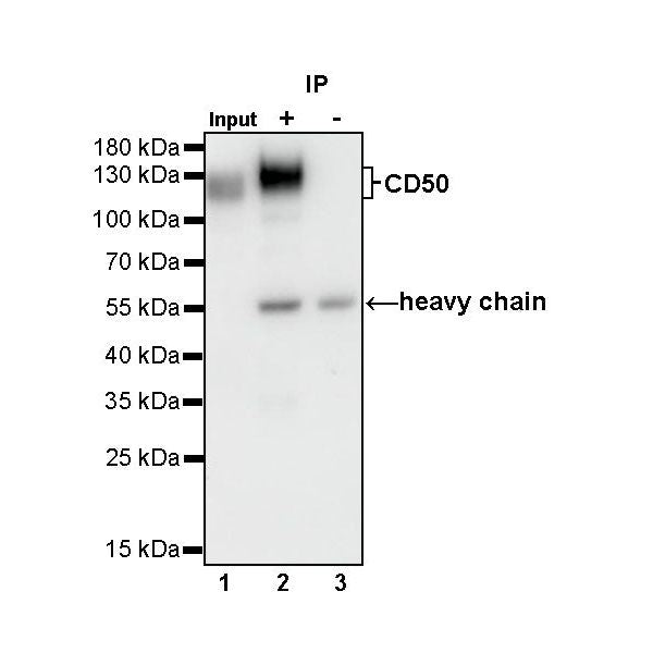
IP
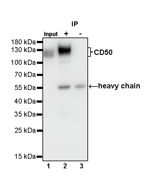
CD50 Rabbit mAb at 1/50 dilution (1 µg) immunoprecipitating CD50 in 0.4 mg Ramos whole cell lysate.
Western blot was performed on the immunoprecipitate using CD50 Rabbit mAb at 1/1000 dilution.
Secondary antibody (HRP) for IP was used at 1/400 dilution.
Lane 1: Ramos whole cell lysate 20 µg (Input)
Lane 2: CD50 Rabbit mAb IP in Ramos whole cell lysate
Lane 3: Rabbit monoclonal IgG IP in Ramos whole cell lysate
Predicted MW: 60 kDa
Observed MW: 110~130 kDa
(This blot was developed with high sensitivity substrate)
Immunohistochemistry
IHC shows positive staining in paraffin-embedded human tonsil. Anti-CD50 antibody was used at 1/500 dilution, followed by a HRP Polymer for Mouse & Rabbit IgG (ready to use). Counterstained with hematoxylin. Heat mediated antigen retrieval with Tris/EDTA buffer pH9.0 was performed before commencing with IHC staining protocol.
IHC shows positive staining in paraffin-embedded human colon. Anti-CD50 antibody was used at 1/500 dilution, followed by a HRP Polymer for Mouse & Rabbit IgG (ready to use). Counterstained with hematoxylin. Heat mediated antigen retrieval with Tris/EDTA buffer pH9.0 was performed before commencing with IHC staining protocol.
IHC shows positive staining in paraffin-embedded human liver. Anti-CD50 antibody was used at 1/500 dilution, followed by a HRP Polymer for Mouse & Rabbit IgG (ready to use). Counterstained with hematoxylin. Heat mediated antigen retrieval with Tris/EDTA buffer pH9.0 was performed before commencing with IHC staining protocol.
IHC shows positive staining in paraffin-embedded human mantle cell lymphoma. Anti-CD50 antibody was used at 1/500 dilution, followed by a HRP Polymer for Mouse & Rabbit IgG (ready to use). Counterstained with hematoxylin. Heat mediated antigen retrieval with Tris/EDTA buffer pH9.0 was performed before commencing with IHC staining protocol.
IHC shows positive staining in paraffin-embedded human diffuse large B-cell lymphoma. Anti-CD50 antibody was used at 1/500 dilution, followed by a HRP Polymer for Mouse & Rabbit IgG (ready to use). Counterstained with hematoxylin. Heat mediated antigen retrieval with Tris/EDTA buffer pH9.0 was performed before commencing with IHC staining protocol.
IHC shows positive staining in paraffin-embedded human Hodgkin’s lymphoma. Anti-CD50 antibody was used at 1/500 dilution, followed by a HRP Polymer for Mouse & Rabbit IgG (ready to use). Counterstained with hematoxylin. Heat mediated antigen retrieval with Tris/EDTA buffer pH9.0 was performed before commencing with IHC staining protocol.
IHC shows positive staining in paraffin-embedded human anaplastic large cell lymphoma. Anti-CD50 antibody was used at 1/500 dilution, followed by a HRP Polymer for Mouse & Rabbit IgG (ready to use). Counterstained with hematoxylin. Heat mediated antigen retrieval with Tris/EDTA buffer pH9.0 was performed before commencing with IHC staining protocol.