WB result of BID Rabbit mAb
Primary antibody: BID Rabbit mAb at 1/1000 dilution
Lane 1: Jurkat whole cell lysate 20 µg
Lane 2: THP-1 whole cell lysate 20 µg
Lane 3: HeLa whole cell lysate 20 µg
Lane 4: MCF7 whole cell lysate 20 µg
Secondary antibody: Goat Anti-Rabbit IgG, (H+L), HRP conjugated at 1/10000 dilution
Predicted MW: 22 kDa
Observed MW: 26 kDa
BID Recombinant Rabbit mAb (S-R300)
BID Recombinant Rabbit mAb (S-R300)
Price:
Regular price
$100 USD
Regular price
Sale price
$100 USD
Unit price
per
For shipping services or bulk orders, you may request a quotation.
Secure checkout with

View full details
Product Details
Product Details
Product Specification
| Host | Rabbit |
| Antigen | BID |
| Synonyms | BH3-interacting domain death agonist, p22 BID (BID) |
| Location | Cytoplasm |
| Accession | P55957 |
| Clone Number | S-R300 |
| Antibody Type | Recombinant mAb |
| Isotype | IgG |
| Application | WB, IHC-P, IP |
| Reactivity | Hu, Ms, Rt |
| Purification | Protein A |
| Concentration | 0.5 mg/ml |
| Conjugation | Unconjugated |
| Physical Appearance | Liquid |
| Storage Buffer | PBS, 40% Glycerol, 0.05%BSA, 0.03% Proclin 300 |
| Stability & Storage | 12 months from date of receipt / reconstitution, -20°C as supplied |
Dilution
| application | dilution | species |
| WB | 1:1000 | null |
| IHC | 1:2000 | null |
| IP | 1:50 | null |
Background
BID is a pro-apoptotic Bcl-2 protein containing only the BH3 domain. In response to apoptotic signaling, BID interacts with another Bcl-2 family protein, Bax, leading to the insertion of Bax into organelle membranes, primarily the outer mitochondrial membrane. The expression of BID is upregulated by the tumor suppressor p53, and BID has been shown to be involved in p53-mediated apoptosis.
Picture
Picture
Western Blot
WB result of BID Rabbit mAb
Primary antibody: BID Rabbit mAb at 1/1000 dilution
Lane 1: Raw 264.7 whole cell lysate 20 µg
Secondary antibody: Goat Anti-Rabbit IgG, (H+L), HRP conjugated at 1/10000 dilution
Predicted MW: 22 kDa
Observed MW: 26 kDa
WB result of BID Rabbit mAb
Primary antibody: BID Rabbit mAb at 1/1000 dilution
Lane 1: PC-12 whole cell lysate 20 µg
Secondary antibody: Goat Anti-Rabbit IgG, (H+L), HRP conjugated at 1/10000 dilution
Predicted MW: 22 kDa
Observed MW: 26 kDa
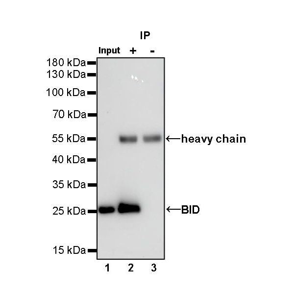
IP
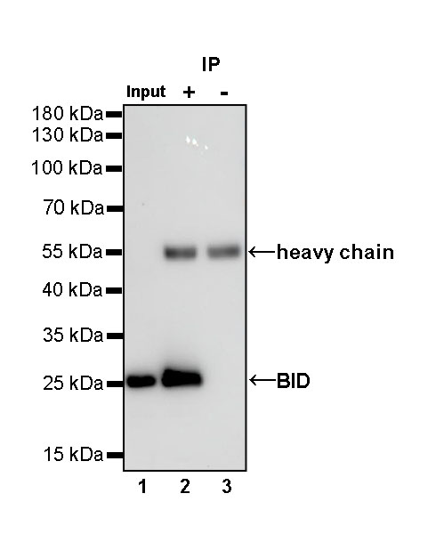
BID Rabbit mAb at 1/50 dilution (1 µg) immunoprecipitating BID in 0.4 mg Jurkat whole cell lysate.
Western blot was performed on the immunoprecipitate using BID Rabbit mAb at 1/1000 dilution.
Secondary antibody (HRP) for IP was used at 1/400 dilution.
Lane 1: Jurkat whole cell lysate 20 µg (Input)
Lane 2: BID Rabbit mAb IP in Jurkat whole cell lysate
Lane 3: Rabbit monoclonal IgG IP in Jurkat whole cell lysate
Predicted MW: 22 kDa
Observed MW: 26 kDa
Immunohistochemistry
IHC shows positive staining in paraffin-embedded human cerebral cortex. Anti-BID antibody was used at 1/2000 dilution, followed by a HRP Polymer for Mouse & Rabbit IgG (ready to use). Counterstained with hematoxylin. Heat mediated antigen retrieval with Tris/EDTA buffer pH9.0 was performed before commencing with IHC staining protocol.
IHC shows positive staining in paraffin-embedded mouse cerebral cortex. Anti-BID antibody was used at 1/2000 dilution, followed by a HRP Polymer for Mouse & Rabbit IgG (ready to use). Counterstained with hematoxylin. Heat mediated antigen retrieval with Tris/EDTA buffer pH9.0 was performed before commencing with IHC staining protocol.
IHC shows positive staining in paraffin-embedded mouse liver. Anti-BID antibody was used at 1/2000 dilution, followed by a HRP Polymer for Mouse & Rabbit IgG (ready to use). Counterstained with hematoxylin. Heat mediated antigen retrieval with Tris/EDTA buffer pH9.0 was performed before commencing with IHC staining protocol.
IHC shows positive staining in paraffin-embedded rat cerebral cortex. Anti-BID antibody was used at 1/2000 dilution, followed by a HRP Polymer for Mouse & Rabbit IgG (ready to use). Counterstained with hematoxylin. Heat mediated antigen retrieval with Tris/EDTA buffer pH9.0 was performed before commencing with IHC staining protocol.