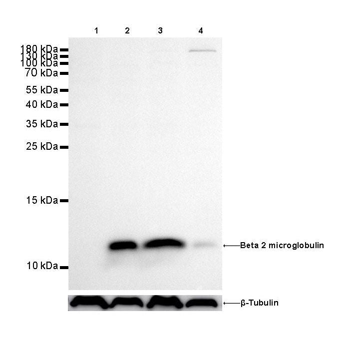
WB result of beta 2 microglobulin Rabbit mAb Primary antibody: beta 2 microglobulin Rabbit mAb at 1/500 dilution
Lane 1: Daudi whole cell lysate 20 µg
Lane 2: Hela whole cell lysate 20 µg
Lane 3: Jurkat whole cell lysate 20 µg
Lane 4: HepG2 whole cell lysate 20 µg
Negative control: Daudi whole cell lysate Secondary antibody: Goat Anti-Rabbit IgG, (H+L), HRP conjugated at 1/10000 dilution
Predicted MW: 12 kDa
Observed MW: 12 kDa
Exposure time: 180s
Beta 2 microglobulin Recombinant rabbit mAb (SDT-096-100)
Beta 2 microglobulin Recombinant rabbit mAb (SDT-096-100)
Price:
Regular price
$100 USD
Regular price
Sale price
$100 USD
Unit price
per
For shipping services or bulk orders, you may request a quotation.
Secure checkout with

View full details
Product Details
Product Details
Product Specification
| Host | Rabbit |
| Antigen | Beta 2 microglobulin |
| Synonyms | Beta-2-microglobulin form pI 5.3, B2M |
| Immunogen | Recombinant Protein |
| Location | Secreted, Cell Surface |
| Accession | P61769 |
| Clone Number | SDT-096-100 |
| Antibody Type | Rabbit mAb |
| Application | WB, IHC-P |
| Reactivity | Hu, Ms |
| Predicted Reactivity | Or |
| Purification | Protein A |
| Concentration | 0.25mg/ml |
| Physical Appearance | Liquid |
| Storage Buffer | PBS, 40% Glycerol, 0.05%BSA, 0.03% Proclin 300 |
| Stability & Storage | 12 months from date of receipt / reconstitution, -20 °C as supplied |
Dilution
| application | dilution | species |
| IHC-P | 1:1000 | |
| WB | 1:500 |
Background
β2 microglobulin (B2M) is a component of MHC class I molecules. MHC class I molecules have α1, α2, and α3 proteins which are present on all nucleated cells (excluding red blood cells). In humans, the β2 microglobulin protein is encoded by the B2M gene. An additional function is association with the HFE protein, together regulating the expression of hepcidin in the liver which targets the iron transporter ferroportin on the basolateral membrane of enterocytes and cell membrane of macrophages for degradation resulting in decreased iron uptake from food and decreased iron release from recycled red blood cells in the MPS (mononuclear phagocyte system) respectively. Loss of this function causes iron excess and hemochromatosis. In patients on long-term hemodialysis, it can aggregate into amyloid fibers that deposit in joint spaces, a disease, known as dialysis-related amyloidosis. Levels of β2 microglobulin can be elevated in multiple myeloma and lymphoma, though in these cases primary amyloidosis (amyloid light chain) and secondary amyloidosis (amyloid associated protein) are more common.[clarification needed] The normal value of β2 microglobulin is < 2 mg/L.
Picture
Picture
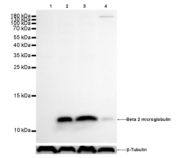
Western Blot
Immunohistochemistry

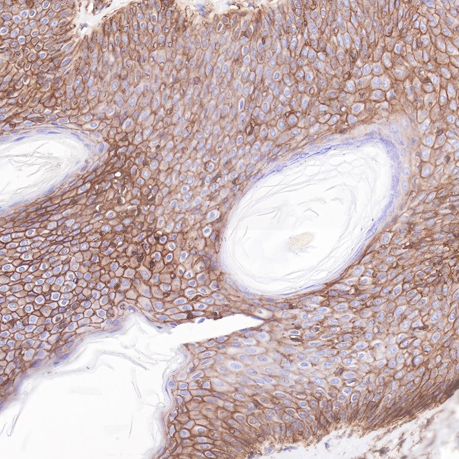
IHC shows positive staining in paraffin-embedded human skin.
Anti-Beta 2 microglobulin antibody was used at 1/1000 dilution, followed by a Goat Anti-Rabbit IgG H&L (HRP) ready to use.
Counterstained with hematoxylin.
Heat mediated antigen retrieval with Tris/EDTA buffer pH9.0 was performed before commencing with IHC staining protocol.
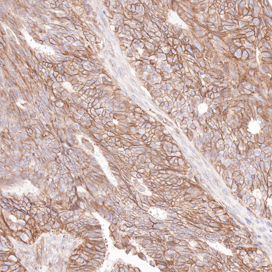
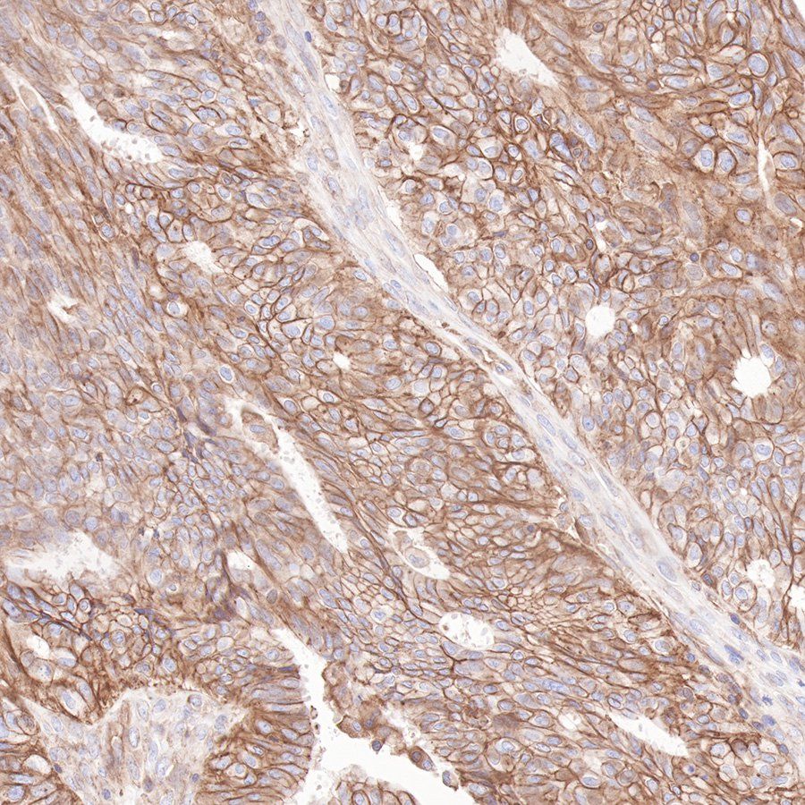
IHC shows positive staining in paraffin-embedded human ovarian cancer.
Anti-Beta 2 microglobulin antibody was used at 1/1000 dilution, followed by a Goat Anti-Rabbit IgG H&L (HRP) ready to use. Counterstained with hematoxylin.
Heat mediated antigen retrieval with Tris/EDTA buffer pH9.0 was performed before commencing with IHC staining protocol.
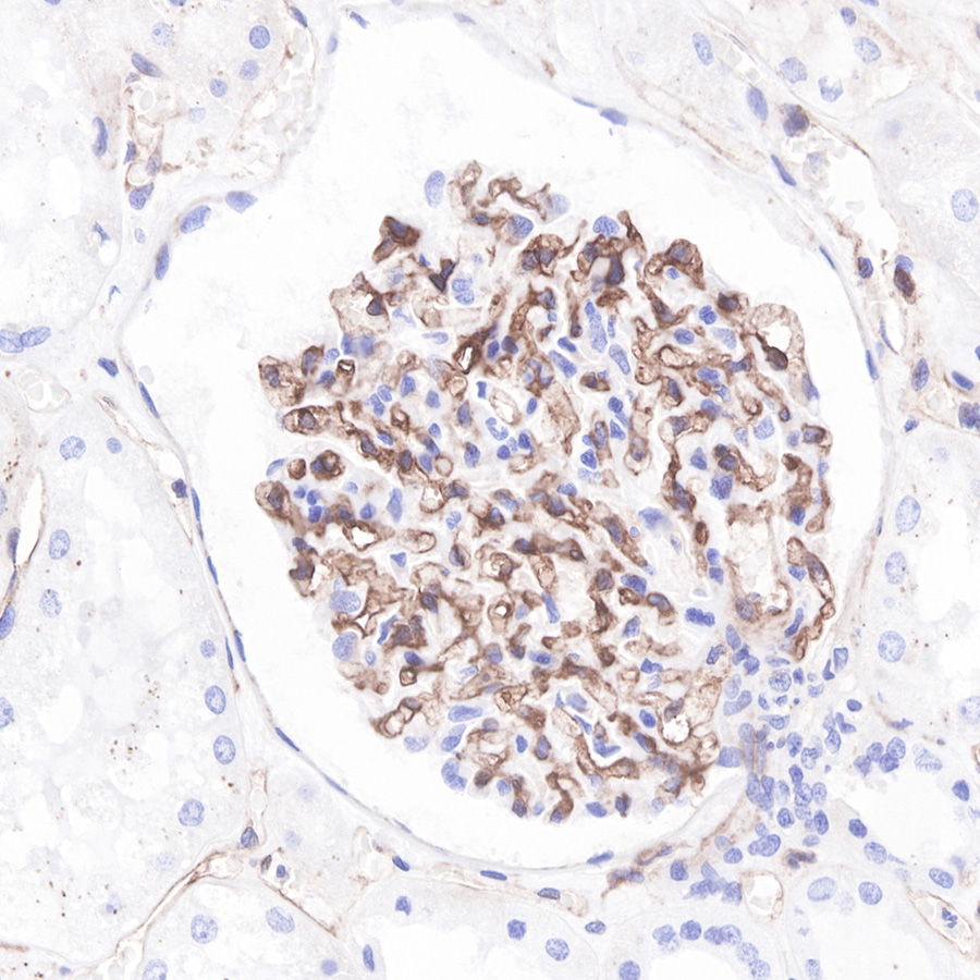
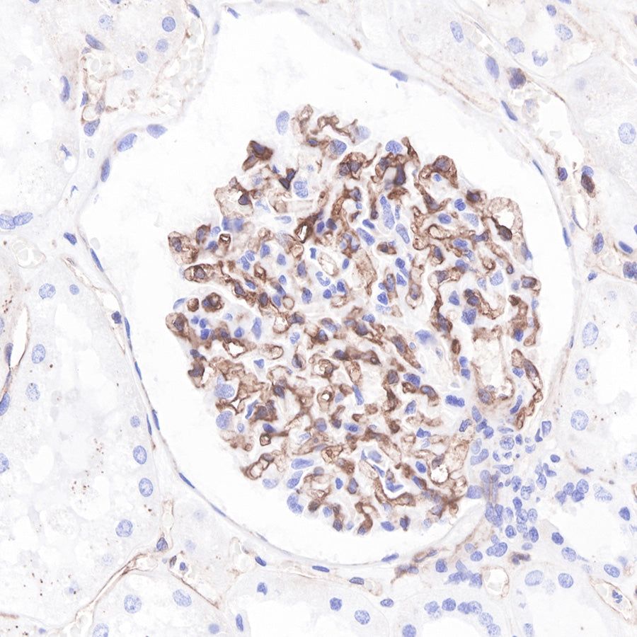
IHC shows positive staining in paraffin-embedded human kidney. Anti-Beta 2 microglobulin antibody was used at 1/1000 dilution, followed by a Goat Anti-Rabbit IgG H&L (HRP) ready to use.
Counterstained with hematoxylin.
Heat mediated antigen retrieval with Tris/EDTA buffer pH9.0 was performed before commencing with IHC staining protocol.
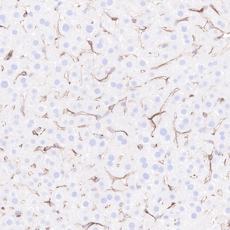
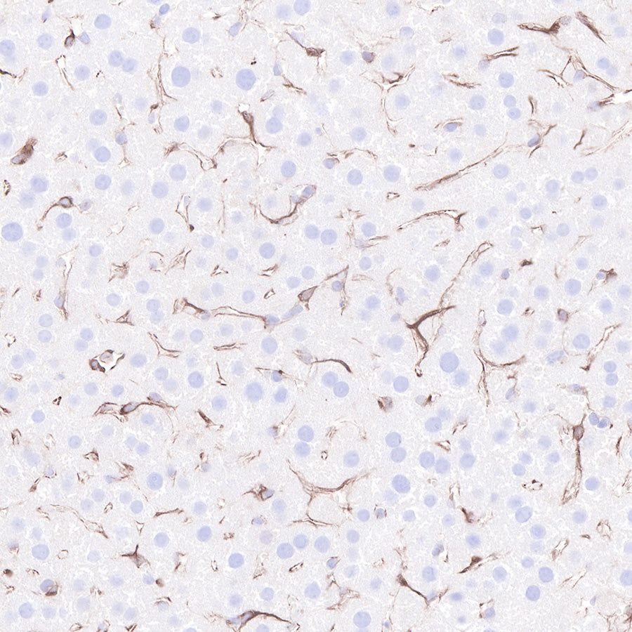
IHC shows positive staining in paraffin-embedded mouse liver.
Anti-Beta 2 microglobulin antibody was used at 1/1000 dilution, followed by a Goat Anti-Rabbit IgG H&L (HRP) ready to use.
Counterstained with hematoxylin.
Heat mediated antigen retrieval with Tris/EDTA buffer pH9.0 was performed before commencing with IHC staining protocol.