
Mouse Total IgG Detection Kit [Wide Range] (HICA)
Mouse Total IgG Detection Kit [Wide Range] (HICA)
Product Details
Product Details
Product Specification
| Stability & Storage | Store at 2~8°C protected from light for 12 months; |
Background
This kit is used for the quantitative detection of mouse immunoglobulin G (mIgG) concentration in buffer solutions, cell culture supernatants, or serum samples.
Mouse immunoglobulin G (mIgG) consists of two heavy chains and two light chains. Based on differences in the constant region of the heavy chains, it can be classified into four main subclasses: IgG1, IgG2a, IgG2b, and IgG3.
Different IgG subclasses exhibit distinct structural and functional characteristics, thereby playing varied roles in immune processes such as antigen recognition, complement activation, and binding to cell surface Fc receptors. Among them, IgG2a and IgG2b typically exhibit strong complement activation capabilities and effectively mediate ADCC (antibody-dependent cell-mediated cytotoxicity) and phagocytosis. IgG1, while relatively weaker in complement activation, primarily functions through Fc receptor-mediated opsonization. IgG3 has been less studied in terms of functionality but is known to play a critical role in immune responses to certain polysaccharide antigens. Clarifying the functional differences among mouse IgG subclasses is of great significance for deepening the understanding of immune response mechanisms, constructing relevant disease models, and evaluating the in vivo effects of therapeutic antibodies.
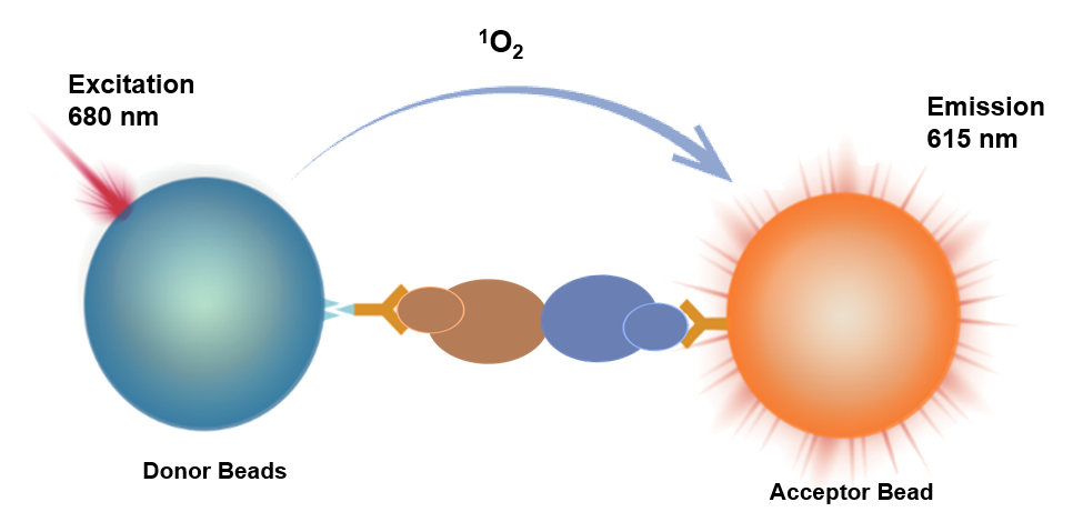
This kit employs a homogeneous luminescence method in a competitive mode to detect mIgG concentration. The homogeneous luminescence method is an immunoassay based on energy transfer between donor and acceptor beads at close proximity.
It uses acceptor beads conjugated with anti-mIgG antibodies (R1), biotin-labeled mIgG (R2), and donor beads conjugated with streptavidin (R3) to determine the concentration of total mouse IgG.
When the test sample contains no mIgG, R2 and R1 form an immune complex with the sample, and the biotin-labeled mIgG binds to the acceptor beads. This complex then binds to the donor beads (R3) to form a luminescent complex. At this point, the distance between the two types of beads is less than 200 nm. Upon excitation light irradiation, the donor beads generate singlet oxygen, which diffuses to the acceptor beads, causing the acceptor beads to emit corresponding luminescence.
Conversely, when the test sample contains mIgG, the mIgG in the sample competitively binds to R1, forming an immune complex. After adding R3, the formation of luminescent complexes between donor and acceptor beads decreases, resulting in a corresponding reduction in the luminescent signal from the acceptor beads upon laser irradiation.
By collecting the luminescent signal with the instrument's photosensitive components and fitting a standard curve using calibrators, the concentration of mIgG in the sample can be calculated.
Components
Name |
Key Component |
Quantity |
mIgGDetection ReagentR1 |
Acceptor Microspheres Conjugated with Anti-Mouse IgG Antibody |
2mL/bottle×1 |
mIgGDetection ReagentR2 |
Biotinylated Mouse IgG |
2mL/bottle×1 |
mIgGDetection ReagentR3 |
Donor Microspheres Conjugated with Streptavidin |
5mL/bottle×1 |
mIgG Calibrator |
Mouse IgG |
100μg,Lyophilized Powder |
mIgG Calibrator Buffer |
BSA |
2mL/bottle×1 |
Protocol
Required Materials and Instruments Not Provided with This Product:
•Luminescent plate/strip
•Multifunctional microplate reader with Alpha module
[Sample Requirements]
•Cell supernatant must be centrifuged at 1000 ×g for 10 minutes to remove particles and polymers.
•If the sample concentration exceeds the detection limit, dilution is recommended before testing.
[Reference Detection Protocol]
Detection Steps |
Detection Protocol (37°C) |
Detection Protocol (Room Temperature) |
Step 1: |
2μL calibrator/sample + 4μL R2* + 4μL R1 + 10μL R3,protected from light/green light |
2μL calibrator/sample + 4μL R2* + 4μL R1 + 10μL R3,protected from light/green light |
Step 2: |
Incubate at 37°C for 30 minutes,protected from light/green light |
Incubate at room temperature for 60 minutes,protected from light/green light |
Reading |
Instrument reading |
Instrument reading |
* This kit uses a competitive assay. Detection reagent R2 reacts directly with R1, so they cannot be pre-mixed. Follow the protocol strictly and pay attention to the order of addition!
[Calibrator Gradient Sample Preparation]
Use the same matrix as the test samples to reconstitute and prepare the calibrator gradient samples. For example, if the test sample is cell culture supernatant, use the same culture medium without cells to reconstitute and prepare the calibrator gradient samples.
Reconstitute the lyophilized calibrator with 100μL of matrix, then dilute the mIgG calibrator with the matrix. The recommended dilution scheme is as follows:
Gradient |
Concentration (μg/mL) |
mIgG High Concentration |
Matrix (μL) |
C8 |
1000 |
/ |
/ |
C7 |
300 |
21μL C8 |
49 |
C6 |
100 |
7μL C8 |
63 |
C5 |
30 |
7μL C7 |
63 |
C4 |
10 |
7μL C6 |
63 |
C3 |
3 |
7μL C5 |
63 |
C2 |
1 |
7μL C4 |
63 |
C1 |
0 |
— |
63 |
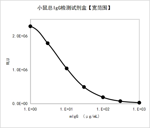
Example of a complete standard curve:
[Performance Parameters]
•Limit of Blank (LoB): Measure calibrator C1 20 times, calculate the mean signal and SD. The concentration corresponding to the mean signal - 2×SD is the LoB.
Detection Protocol |
Matrix |
LoB (μg/mL) |
Protocol 1 |
Calibrator buffer |
0.74 |
Protocol 2 |
Calibrator buffer |
0.64 |
•Dynamic Range: 0~1000 μg/mL.
•Repeatability: Test high and low concentration samples 10 times and calculate the CV of concentration.
Detection Protocol |
Matrix |
Repeatability |
|
Low Concentration |
High Concentration |
||
Protocol 1 |
Calibrator buffer |
2.31% |
2.71% |
Protocol 2 |
Calibrator buffer |
4.88% |
2.23% |
•Accuracy: Test accuracy samples and calculate the deviation from the target value.
Detection Protocol |
Matrix |
Deviation |
|
Low Concentration |
High Concentration |
||
Protocol 1 |
Calibrator buffer |
6.85% |
8.25% |
Protocol 2 |
Calibrator buffer |
-7.77% |
-9.03% |
•Specificity: Dilute the following cross-reactive substances to 100 μg/mL using calibrator buffer and test the cross-reactivity rate.
Detection Protocol |
Cross-Reactive Substance |
Cross-Reactivity Rate |
Protocol 1 |
Human IgG |
8.20% |
Rabbit IgG |
0.16% |
|
Porcine IgG |
0.23% |
Guidelines
The detection reagent R3 is light-sensitive. Avoid exposure to light during use, and it is recommended to perform sample addition and incubation under green light (illuminance < 100 LUX).
It is recommended to recalibrate for each detection, with 2-3 replicate wells for each standard concentration point.
Four-parameter (weighting 1/Y²) or cubic spline fitting is recommended for calculation.
Pay attention to the requirements for incubation temperature and duration.
For 37°C incubation, it is recommended to use the HiLA homogeneous luminescence analyzer.
Components from different reagent kit batches should not be mixed.
![Mouse Total IgG Detection Kit [Wide Range] (HICA)](http://www.antbioinc.com/cdn/shop/files/AntBioImage_56e1c74d-5f0f-47b4-b215-7749a96cdb77.png?v=1780633741&width=1445)