Human Total IgG Kit (Wide Range) (HICA)
Human Total IgG Kit (Wide Range) (HICA)
Product Details
Product Details
Product Specification
| Stability & Storage | Store at 2~8°C, protected from light for 12 months |
Background
This product is used for the quantitative detection of the total concentration of human immunoglobulin G (hIgG) in buffer solutions, cell culture supernatants, or serum samples.
Human immunoglobulin G (hIgG) is composed of two heavy chains and two light chains. Based on differences in the constant region of the heavy chains, it can be classified into four subclasses: IgG1, IgG2, IgG3, and IgG4, with IgG1 having the highest expression level and IgG4 the lowest.
Structural and functional differences among IgG subclasses directly influence their roles in immune processes such as antigen recognition, complement activation, and binding to cell surface receptors. Studies have shown that changes in the concentrations of IgG subclasses in serum are closely associated with various disease states¹. Functionally, IgG1 and IgG3 exhibit strong complement activation capabilities, promoting phagocytosis and serving as critical effector molecules in the body's defense against infections. In contrast, IgG4 primarily participates in anti-inflammatory responses and the maintenance of immune tolerance, and its abnormal elevation may be linked to tumor immune evasion or the development of IgG4-related diseases.
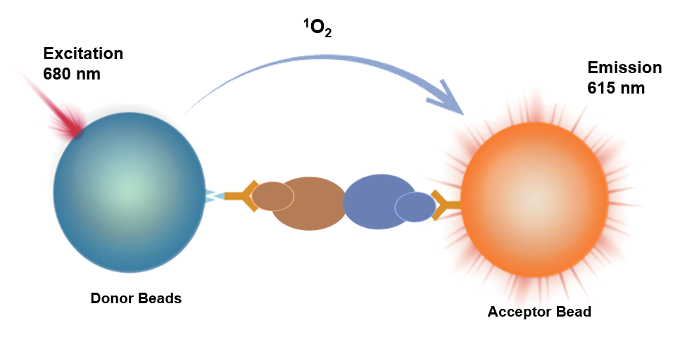
This kit employs a homogeneous luminescence-based competitive assay for the detection of hIgG concentration. Homogeneous chemiluminescence is an immunoassay method based on energy transfer between donor and acceptor microspheres to produce luminescence.
It utilizes acceptor microspheres conjugated with anti-hIgG antibodies (R1), biotin-labeled hIgG (R2), and donor microspheres conjugated with streptavidin (R3) to measure the total concentration of human IgG.
When the test sample contains no hIgG, after mixing R2 and R1 with the sample, the biotin-labeled hIgG forms an immune complex with the acceptor microspheres. Upon addition of R3, the donor microspheres bind to form a luminescent complex, where the distance between the two microspheres is less than 200 nm. Upon excitation, the donor microspheres generate singlet oxygen, which diffuses to the acceptor microspheres, and the acceptor microspheres emit corresponding light upon receiving the energy.
Conversely, when the test sample contains hIgG, the hIgG in the sample competitively binds with R1 to form an immune complex, reducing the formation of the luminescent complex with donor and acceptor microspheres upon addition of R3. Upon laser excitation, the light signal produced by the acceptor microspheres decreases accordingly.
The light signal is collected by the instrument's photosensitive element, and the concentration of hIgG in the sample can be calculated using a standard curve fitted with calibrators.
Components
Name |
Main Active Ingredient |
Content |
hIgG Detection ReagentR1 |
Anti-hIgG Fc antibody-conjugated acceptor beads |
2mL/vial ×1 |
hIgG Detection ReagentR2 |
Biotin-labeled hIgG |
2mL/vial ×1 |
hIgG Detection ReagentR3 |
Streptavidin-conjugated donor beads |
5mL/vial ×1 |
hIgG Calibrator |
hIgG protein |
50μL,0.5mg |
Protocol
Materials and Instruments Required but Not Provided with this Product:
•Luminescence plates/tubes
•Multimode microplate reader equipped with Alpha module
[Sample Requirements]
•Cell culture supernatants must be centrifuged at 1000 ×g for 10 minutes to remove particles and polymers.
•If the concentration of the test sample exceeds the upper limit of detection, dilution prior to testing is recommended.
[Assay Procedure for Reference]
Assay Procedure |
Assay Procedure (37°C) |
Assay Procedure (Room Temperature) |
Step 1: |
2μL Calibrator/Sample + 4μL R2* + 4μL R1 + 10μL R3,Avoid Light/Green Light |
2μL Calibrator/Sample + 4μL R2* + 4μL R1 + 10μL R3,Avoid Light/Green Light |
Step 2: |
Incubate at 37°C for 30 minutes,Avoid Light/Green Light |
Incubate at room temperature for 60 minutes,Avoid Light/Green Light |
Read |
Instrument Read |
Instrument Read |
* This kit utilizes a competitive assay format. Detection reagent R2 reacts directly with R1; they must not be pre-mixed before addition. Pay attention to the addition sequence and strictly follow the procedure!
[Calibrator Gradient Sample Preparation]
Prepare calibrator gradient samples using the same matrix as the test samples. For example, if the test samples are cell culture supernatants, use cell-free culture medium to prepare the calibrator gradient samples.
Retrieve the calibrators, then dilute the hIgG calibrators using the matrix. The recommended dilution scheme is as follows:
Gradient |
Concentration (μg/mL) |
High Concentration hIgG |
Matrix (μL) |
C8 |
1000 |
20μL Calibrator |
180 |
C7 |
300 |
60μL C8 |
140 |
C6 |
100 |
60μL C7 |
120 |
C5 |
30 |
60μL C6 |
140 |
C4 |
10 |
60μL C5 |
120 |
C3 |
3 |
60μL C4 |
140 |
C2 |
1 |
60μL C3 |
120 |
C1 |
0 |
— |
120 |
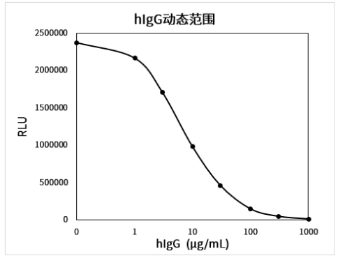
Example of Complete Standard Curve (Dilution Matrix: 20 mM PBS):
[Performance Parameters]
•Limit of Blank (LOB): Test Calibrator C1 in duplicate 20 times, calculate the mean signal and SD. Use the standard curve to calculate the concentration corresponding to Mean Signal - 2×SD, which defines the LOB.
Assay Procedure |
Matrix |
Limit of Blank (μg/mL) |
Assay Procedure 1 |
PBS Buffer |
0.58 |
Assay Procedure 2 |
PBS Buffer |
0.30 |
•Dynamic Range: 0–1000 μg/mL.
•Repeatability: High (~500 μg/mL) and low (~100 μg/mL) concentration samples were tested 10 times in replicate, and the concentration CV was calculated.
Assay Procedure |
Matrix |
Repeatability CV |
|
Low Concentration |
High Concentration |
||
Assay Procedure 1 |
PBS Buffer |
3.9% |
3.3% |
Assay Procedure 2 |
PBS Buffer |
4.7% |
4.5% |
•Accuracy: Test accuracy control samples and calculate the deviation from the target value.
Assay Procedure |
Matrix |
Accuracy Samples |
|
Low Concentration |
High Concentration |
||
Assay Procedure 1 |
PBS Buffer |
2.1% |
-4.5% |
Assay Procedure 2 |
PBS Buffer |
-3.3% |
-7.7% |
•Specificity: Dilute the following cross-reactants to 100 μg/mL using PBS buffer and test the cross-reactivity rate.
Assay Procedure |
Cross-reactant |
Cross-reactivity Rate |
Assay Procedure 1 |
Mouse IgG |
0.01% |
Rabbit IgG |
2.63% |
|
Pig IgG |
0.51% |
Guidelines
Detection reagent R3 is light-sensitive. Avoid exposure to light during use, and it is recommended to perform sample addition and incubation under green light (illuminance < 100 LUX).
It is recommended to recalibrate for each detection, with 2~3 replicates for each concentration point of the standard.
Four-parameter (weight 1/Y²) or cubic spline fitting is recommended for calculation.
Pay attention to the requirements for incubation temperature and duration.
For 37℃ incubation, it is recommended to use the HiLA homogeneous luminescence analyzer.
Components from different reagent kit batches should not be mixed.