
Human IgG1 Detection Kit [Wide Range] (HICA)
Human IgG1 Detection Kit [Wide Range] (HICA)
Product Details
Product Details
Product Specification
| Stability & Storage | Store at 2-8°C away from light; product shelf life is 12 months. |
Background
This product is used for the quantitative detection of human immunoglobulin G1 (hIgG1) concentration in buffer solutions, cell culture supernatants, or serum samples.
Human immunoglobulin G (hIgG) consists of two heavy chains and two light chains. Based on differences in the constant regions of the heavy chains, it can be further classified into four subclasses: IgG1, IgG2, IgG3, and IgG4, with IgG1 being the most abundant and IgG4 the least.
Different IgG subclasses exhibit variations in structure and function, thereby playing distinct roles in immune processes such as antigen recognition, complement activation, and binding to cell surface receptors. Numerous studies have shown that deviations in serum concentrations of IgG subclasses from normal reference ranges are often closely associated with various disease states. For example, IgG1 and IgG3 have strong complement activation capabilities, promoting phagocytosis and serving as crucial effector molecules in the body's defense against infections. In contrast, IgG4 is related to anti-inflammatory responses and immune tolerance, and its abnormal elevation may be linked to tumor immune evasion or the development of IgG4-related diseases.
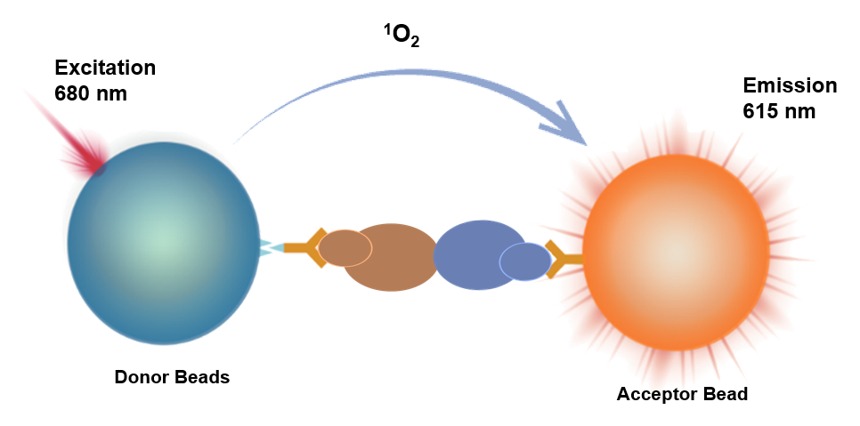
This kit employs a homogeneous luminescence-based competitive assay for the detection of hIgG1 concentration. Homogeneous chemiluminescence is an immunoassay method based on energy transfer between donor and acceptor beads in close proximity.
It utilizes acceptor beads conjugated with anti-hIgG antibodies (R1), biotin-labeled hIgG1 (R2), and donor beads conjugated with streptavidin (R3) to measure the concentration of human IgG1.
When the test sample contains no hIgG1, R2 and R1 mix with the sample, and the biotin-labeled hIgG1 forms an immune complex with the acceptor beads. This complex then binds to the donor beads of R3, forming a luminescent complex. In this case, the distance between the two types of beads is less than 200 nm. Upon excitation, the donor beads generate singlet oxygen, which diffuses to the acceptor beads, causing them to emit corresponding light signals.
Conversely, when the test sample contains hIgG1, the hIgG1 in the sample competitively binds with R1 to form immune complexes, reducing the formation of luminescent complexes with donor and acceptor beads upon the addition of R3. Upon laser excitation, the light signals produced by the acceptor beads decrease accordingly.
By collecting the light signals with the instrument's photosensitive elements and fitting a standard curve using calibrators, the concentration of hIgG1 in the sample can be calculated.
Components
Name |
Main Active Ingredient |
Content |
hIgG1 Detection Reagent R1 |
Anti-hIgG Fc antibody-conjugated acceptor beads |
2mL/vial×1 |
hIgG1 Detection Reagent R2 |
Biotin-labeled hIgG1 |
2mL/vial×1 |
hIgG1 Detection Reagent R3 |
Streptavidin-conjugated donor beads |
5mL/vial×1 |
hIgG1 Calibrator |
hIgG1 protein |
50μL,0.5mg |
Protocol
Required Materials and Instruments Not Provided with This Product:
Luminescence Plate/Strip
Multimode Microplate Reader equipped with an Alpha module
[Sample Requirements]
•Cell supernatants must be centrifuged at 1000 ×g for 10 minutes to remove particles and polymers.
•If the concentration of the test sample exceeds the upper limit of detection, dilution prior to testing is recommended.
[Assay Procedure Reference]
Assay Procedure |
Assay Procedure (37°C) |
Assay Procedure (Room Temperature) |
Step 1: |
2μL Calibrator/Sample + 4μL R2* + 4μL R1 + 10μL R3,Protect from light/Green light |
2μL Calibrator/Sample + 4μL R2* + 4μL R1 + 10μL R3,Protect from light/Green light |
Step 2: |
Incubate at 37°C for 30 minutes,Protect from light/Green light |
Incubate at room temperature for 60 minutes,Protect from light/Green light |
Reading |
Instrument reading |
Instrument reading |
* This kit uses a competitive assay format. Reagent R2 reacts directly with R1; they must not be pre-mixed before addition. Pay attention to the order of addition and strictly follow the procedure!
[Preparation of Calibrator Gradient Samples]
Prepare calibrator gradient samples using the same matrix as the test samples. For example, when testing cell culture supernatants, use cell-free culture medium to prepare the calibrator gradient samples.
Take out the calibrators, then dilute the hIgG1 calibrators using the matrix. The recommended dilution scheme is shown in the table below:
Gradient |
Concentration (μg/mL) |
hIgG1 High Concentration |
Matrix (μL) |
C8 |
1000 |
20μL Calibrator |
180 |
C7 |
300 |
60μL C8 |
140 |
C6 |
100 |
60μL C7 |
120 |
C5 |
30 |
60μL C6 |
140 |
C4 |
10 |
60μL C5 |
120 |
C3 |
3 |
60μL C4 |
140 |
C2 |
1 |
60μL C3 |
120 |
C1 |
0 |
— |
120 |
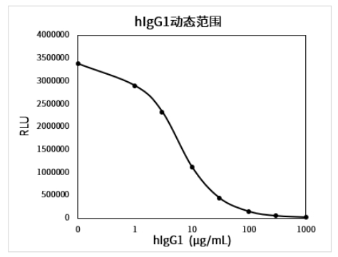
Example of a complete standard curve (dilution matrix is 20mM PBS):
[Performance Parameters]
•Limit of Blank (LOB): Test Calibrator C1 in duplicate 20 times, calculate the mean signal and SD. Use the standard curve to calculate the concentration corresponding to Mean Signal - 2×SD, which is the LOB.
Assay Procedure |
Matrix |
Limit of Blank (μg/mL) |
Assay Procedure 1 |
PBS Buffer |
0.39 |
Assay Procedure 2 |
PBS Buffer |
0.45 |
•Dynamic Range: 0~1000 μg/mL.
•Precision: High (~500 μg/mL) and low (~100 μg/mL) concentration samples were tested in replicate 10 times to calculate the coefficient of variation (CV) for concentration.
Assay Procedure |
Matrix |
Precision CV |
|
Low Concentration |
High Concentration |
||
Assay Procedure 1 |
PBS Buffer |
5.2% |
3.0% |
Assay Procedure 2 |
PBS Buffer |
5.4% |
5.2% |
•Accuracy: Test accuracy samples and calculate the deviation from the target value.
Assay Procedure |
Matrix |
Accuracy Sample |
|
Low Concentration |
High Concentration |
||
Assay Procedure 1 |
PBS Buffer |
6.7% |
2.3% |
Assay Procedure 2 |
PBS Buffer |
3.6% |
2.9% |
•Specificity: Dilute the following cross-reactants to 100 μg/mL using PBS buffer to test the cross-reactivity rate.
Assay Procedure |
Cross-reactant |
Cross-reactivity Rate |
Assay Procedure 1 |
Mouse IgG |
0.00% |
Rabbit IgG |
1.10% |
|
Porcine IgG |
0.13% |
Guidelines
•The detection reagent R3 is light-sensitive. Avoid exposure to light during use, and it is recommended to add samples and incubate under green light (illuminance < 100 LUX).
•It is recommended to recalibrate for each test, with 2–3 replicate wells for each standard concentration.
•Four-parameter (weighting 1/Y²) or cubic spline fitting is recommended for calculation.
•Pay attention to the requirements for incubation temperature and time.
•For 37°C incubation, it is recommended to use the HiLA homogeneous luminescence analyzer.
•Components from different reagent kit batches should not be mixed.
![Human IgG1 Detection Kit [Wide Range] (HICA)](http://www.antbioinc.com/cdn/shop/files/AntBioImage_00d39592-0688-48b0-b8f2-d69935cd8c9f.png?v=1780633741&width=1445)