Human IgG Kit (HICA)
Human IgG Kit (HICA)
Product Details
Product Details
Product Specification
Background
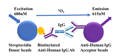
Testing Principle:
This kit employs the Homogeneous Immuno Chemiluminescence Assay (HICA), a highly sensitive homogeneous detection technology based on beads. This technology utilizes two types of beads: donor beads and acceptor beads. When molecules attached to the donor beads interact with molecules attached to the acceptor beads, and the two beads come into sufficiently close proximity (<200 nm), a luminescent signal can be detected.
The photosensitizer on the surface of the donor beads is excited by irradiation with 680 nm laser light, converting surrounding oxygen into singlet oxygen. The singlet oxygen diffuses in the solution and, upon encountering an acceptor bead, triggers a cascade of amplified chemical reactions within it, ultimately resulting in light emission. As illustrated, the streptavidin on the donor bead binds to a biotinylated antibody. The antibody-labeled acceptor bead and the biotinylated antibody both bind to the IgG sample, generating the detection signal. This technology requires no washing steps, is simple to operate, and compared to traditional ELISA, offers a wider detection range, higher sensitivity, and lower background.
Components
Components |
Concentration |
200T |
500T |
5000T |
|---|---|---|---|---|
Streptavidin-labeled Donor Beads |
125× |
8 µL |
20 µL |
200 µL |
Anti-human IgG Antibody-labeled Acceptor Beads |
500× |
2 µL |
5 µL |
50 µL |
Biotin-labeled Anti-human IgG Antibody |
625× |
2 µL |
5 µL |
50 µL |
IgG Standard |
1 mg/mL |
5 µL |
10 µL |
20 µL |
Assay Buffer |
1× |
4 mL |
10 mL |
100 mL |
Protocol
Taking a 384-well shallow well plate as an example
I. Preparation of Working Solutions
Before using donor beads and acceptor beads, ensure the beads are in suspension. If there is slight clumping, resuspend the beads using sonication. Do not allow direct contact between beads and ice cubes. Protect from light during use.
All components should be diluted with 1× Assay Diluent. It is recommended to store and dilute the standard on ice after thawing. The donor beads should be diluted according to the dilution factor into the pre-diluted biotinylated antibody. The volume of the mixed solution of the two reagents per well is 5uL.
1. Anti-human IgG antibody-labeled acceptor beads: After resuspending the acceptor beads, dilute them 500-fold with assay diluent. Use 5uL per well.
2. Biotin-labeled anti-human IgG antibody and streptavidin-labeled donor beads: After diluting the biotinylated antibody with assay diluent, directly dilute the donor beads into the diluted antibody. Use 5uL per well.
3. IgG Standard/Test Samples: Store the standard on ice after thawing. Perform serial dilutions with assay diluent. Use 10uL per well.
Adjust dilutions according to the number of standard curves required for the experiment. For example: dilute 1uL of IgG standard into 249uL of assay diluent to obtain Std 1, then perform 3-fold serial dilutions up to Std 10.
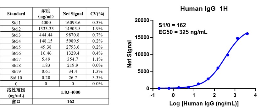
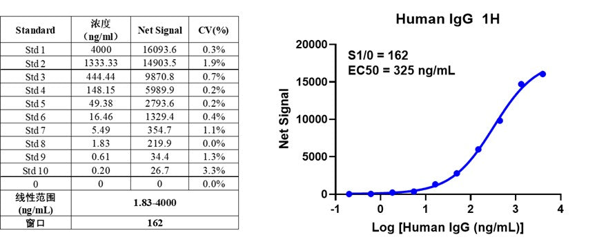
| Standard | Standard Dilution | Working Solution Concentration (ng/mL) |
| Std 1 | 1uL Standard Stock + 249uL Standard Diluent | 4000.00 |
| Std 2 | 10uL Std 1 + 20uL Standard Diluent | 1333.33 |
| Std 3 | 10uL Std 2 + 20uL Standard Diluent | 444.44 |
| Std 4 | 10uL Std 3 + 20uL Standard Diluent | 148.15 |
| Std 5 | 10uL Std 4 + 20uL Standard Diluent | 49.38 |
| Std 6 | 10uL Std 5 + 20uL Standard Diluent | 16.46 |
| Std 7 | 10uL Std 6 + 20uL Standard Diluent | 5.49 |
| Std 8 | 10uL Std 7 + 20uL Standard Diluent | 1.83 |
| Std 9 | 10uL Std 8 + 20uL Standard Diluent | 0.61 |
| Std 10 | 10uL Std 9 + 20uL Standard Diluent | 0.20 |
| 0 | 20uL Standard Diluent | 0.00 |
II. Sample Addition
Once samples, standards, and working solutions of detection reagents are prepared, add them and incubate according to the order specified in the table.
| Component | Test Well | Control Wells | ||
| Negative Control | Donor Bead Control | Acceptor Bead Control | ||
| IgG Standard/Test Sample | 10uL | -- | -- | -- |
| Diluent | -- | 10uL | 15uL | 10uL |
| Anti-human IgG antibody-labeled acceptor beads | 5uL | 5uL | -- | 5uL |
| Step 1: Incubate at room temperature for 1 hour | ||||
| Biotin-labeled anti-human IgG antibody and streptavidin-labeled donor beads | 5uL | 5uL | 5uL | -- |
| Diluent | -- | -- | -- | 5uL |
| Step 2: Incubate at room temperature for 1 hour | ||||
The experiment should include the following control wells:
(1) Buffer Control (Negative Control): To exclude the effect of the buffer on signal values, serving as a background value for calculating the specific signal of the reaction system.
(2) Donor/Acceptor Bead Control Wells: To detect the signal value of isolated donor/acceptor beads in the buffer, used to verify the normal signal value and background of the beads.
III. Detection
Before the experiment, please confirm the functions and configuration of the microplate reader. Perform detection on a compatible homogeneous chemiluminescence microplate reader.
IV. Result Calculation
1. Calculate the net signal ratio of acceptor and donor emission signals for each well
Net signal = [signal (Test Well) - signal (Negative Control Well)] / signal (Negative Control Well)*100
2. Calculate the coefficient of variation (CV%) for replicate wells
Coefficient of Variation CV% = Standard Deviation/Mean*100%
V. Example Data
The following examples are for reference only. Specific experimental systems should be adjusted and optimized according to actual conditions. For each detection, sample concentrations should be calculated using the standard curve generated from the standards run in the same experiment.
Picture
Picture
Bioactivity