Human CTXI ELISA Kit
Human CTXI ELISA Kit
Price:
Regular price
$368 USD
Regular price
Sale price
$368 USD
Unit price
per
For shipping services or bulk orders, you may request a quotation.
Secure checkout with

View full details
Product Details
Product Details
Product Specification
| Usage | Required experimental equipment: 1. Microplate reader (450nm) 2. High-precision pipettes and pipette tips: 0.5-10uL, 5-50uL, 20-200uL, 200-1000uL 3. 37°C incubator 4. Distilled or deionized water Sample handling and requirements: The detection range of the kit is not equivalent to the concentration range of the analyte in the sample. Before the experiment, it is recommended to estimate the analyte concentration in the sample based on relevant literature and conduct preliminary experiments to determine the actual concentration in the sample. If the analyte concentration in the sample is too high or too low, dilute or concentrate the sample appropriately. If the sample type is not listed in the instructions, it is recommended to conduct a preliminary experiment to verify the validity of the test. Serum: Incubate whole blood samples collected in serum separator tubes at room temperature for 2 hours or at 4°C overnight, then centrifuge at 1000×g for 20 minutes. Remove the supernatant for analysis, or store at -20°C or -80°C, but avoid repeated freezing and thawing. Plasma: Collect samples using EDTA or heparin as an anticoagulant and centrifuge at 1000×g for 15 minutes at 2-8°C within 30 minutes of collection. Remove the supernatant for analysis, or store at -20°C or -80°C, but avoid repeated freezing and thawing. Tissue homogenate: Rinse tissue with pre-chilled PBS (0.01M, pH 7.4) to remove residual blood (lysed red blood cells in the homogenate may affect the measurement results). Weigh and mince the tissue. Add the minced tissue to the appropriate volume of PBS (generally a 1:9 weight-to-volume ratio, e.g., 1g of tissue sample to 9mL of PBS. The specific volume can be adjusted according to experimental needs and recorded. It is recommended to add protease inhibitors to the PBS) in a glass homogenizer and grind thoroughly on ice. To further lyse the tissue cells, the homogenate can be sonicated or repeatedly freeze-thawed. Finally, centrifuge the homogenate at 5000×g for 5-10 minutes, and collect the supernatant for analysis. Cell Lysis Buffer: Adherent cells are gently washed with ice-cold PBS, then trypsinized and harvested by centrifugation at 1000×g for 5 minutes. Suspension cells can be harvested directly by centrifugation. The harvested cells are washed three times with ice-cold PBS and resuspended in 150-200µL of PBS per 1×10^6 cells (it is recommended to add protease inhibitors to the PBS; if the cell count is very low, the PBS volume can be reduced appropriately). Disrupt the cells by repeated freeze-thaw cycles or sonication. Centrifuge the extract at 1500×g for 10 minutes at 2-8°C, and remove the supernatant for testing. Cell culture supernatant: Centrifuge at 1000×g for 20 minutes, remove the supernatant, and test immediately. Alternatively, store the supernatant at -20°C or -80°C, but avoid repeated freezing and thawing. Other biological samples: Centrifuge at 1000×g for 20 minutes, remove the supernatant, and test immediately. Sample Appearance: The sample should be clear and transparent, and suspended matter should be removed by centrifugation. Sample Storage: Samples collected for testing within one week can be stored at 4°C. If testing is not possible, aliquot the sample into single-use aliquots and freeze at -20°C (for testing within one month) or -80°C (for testing within six months). Avoid repeated freezing and thawing. Hemolysis of the sample can affect the final test results, so hemolyzed samples are not suitable for this test. Preparation for the test: 1. Remove the reagent kit from the refrigerator 10 minutes in advance and equilibrate to room temperature. 2. Prepare a gradient standard working solution: Add 1 mL of universal diluent to the lyophilized standard. Let stand for 15 minutes to completely dissolve, then gently mix (concentration 10 ng/mL). Then dilute to the following concentrations: 10 ng/mL, 5 ng/mL, 2.5 ng/mL, 1.25 ng/mL, 0.625 ng/mL, 0.3125 ng/mL, 0.15625 ng/mL, and 0 ng/mL. Serial dilution method: Take 7 EP tubes and add 500 μL of universal diluent to each tube. Pipette 500 μL of the 10 ng/mL standard working solution into the first EP tube and mix thoroughly to make a 5 ng/mL standard working solution. Repeat this procedure for subsequent tubes. The last tube serves as a blank well; there is no need to pipette liquid from the penultimate tube. See the figure below for details. 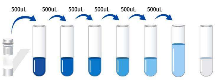 3. Preparation of Biotin-Antibody Working Solution: 15 minutes before use, centrifuge the concentrated biotinylated antibody at 1000×g for 1 minute. Dilute the 100× concentrated biotinylated antibody to a 1× working concentration with universal diluent (e.g., 10 μL concentrate + 990 μL universal diluent). Prepare and use immediately. 4. Preparation of Enzyme Conjugate Working Solution: 15 minutes before use, centrifuge 100 μL of concentrated enzyme conjugate at 1000 × g for 1 minute. Dilute the 100 μL concentrated HRP enzyme conjugate with universal diluent to a 1 μL working concentration (e.g., 10 μL concentrate + 990 μL universal diluent). Prepare freshly for use. 5. Preparation of 1 μL Wash Solution: Dissolve 10 mL of 20 μL Wash Solution in 190 mL of distilled water. (Crystallization may occur in the concentrated wash solution after removal from the refrigerator. This is normal. Allow to stand at room temperature until the crystals have completely dissolved before preparation.) Procedure: 1. After equilibration at room temperature for 10 minutes, remove the desired strips from the aluminum foil bag. Seal the remaining strips in a ziplock bag and return to 4°C. 2. Sample Addition: Add 50 μL of sample or standard of varying concentrations to the appropriate wells. Add 50 μL of universal diluent to the blank wells, followed by 50 μL of Biotin-Antibody Working Solution to each well. Cover with a film sealer and incubate at 37°C for 60 minutes. (Recommendation: Dilute the sample to be tested at least 1x in universal diluent before adding it to the plate to minimize matrix effects. When calculating sample concentration, multiply by the dilution factor. It is recommended to run replicates for all samples and standards.) 3. Wash: Discard the remaining solution and add 300 μL of 1x wash buffer to each well. Let stand for 1 minute, then shake off the wash buffer and pat dry on absorbent paper. Repeat this process three times (a microplate washer can also be used). 5. Add Enzyme Conjugate Working Solution: Add 100 μL of enzyme conjugate working solution to each well, cover with a film sealer, and incubate at 37°C for 30 minutes. 6. Wash: Discard the solution and wash the plate five times according to the procedure in step 4. 7. Add Substrate: Add 90 μL of substrate (TMB) to each well, cover with a film sealer, and incubate at 37°C in the dark for 15 minutes. 8. Add Stop Solution: Remove the plate and add 50 μL of stop solution directly to each well. Immediately measure the OD value of each well at 450 nm. Calculation of Experimental Results: 1. Calculate the average OD value of the standard and sample replicates and subtract the OD value of the blank well as a correction value. Plot a standard curve for the four-parameter logistic function on double-logarithmic graph paper with concentration as the horizontal axis and OD value as the vertical axis. 2. If the sample OD value is higher than the upper limit of the standard curve, it should be diluted appropriately and re-measured and the sample concentration should be multiplied by the corresponding dilution factor when calculating.  |
|||||||||||||||||||||||||||||||||
| Sensitivity | 0.05 ng/mL | |||||||||||||||||||||||||||||||||
| Theory | This kit uses a competitive enzyme-linked immunosorbent assay (ELISA). Sample, standard, biotin-labeled antibody, and HRP conjugate are sequentially added to microwells pre-coated with human type I collagen cross-linked carboxyl-terminal peptide (CTXI) antigen. After incubation and washing, the sample is developed using the substrate TMB. TMB is converted to blue by HRP peroxidase and to yellow by acid. The intensity of the color is negatively correlated with the amount of human type I collagen cross-linked carboxyl-terminal peptide (CTXI) in the sample. The absorbance (OD) is measured at 450 nm using a microplate reader to calculate the sample concentration. | |||||||||||||||||||||||||||||||||
| Source | Human | |||||||||||||||||||||||||||||||||
| Synonym | Human Cross Linked C-Telopeptide Of Type I Collagen ELISA Kit | |||||||||||||||||||||||||||||||||
| Detection Type | Competition Law | |||||||||||||||||||||||||||||||||
| Composition |
|
|||||||||||||||||||||||||||||||||
| Background | CTX-1, also known as the carboxy-terminal cross-linked telopeptide of type 1 collagen or the C-terminal telopeptide of type I collagen, is a marker of osteoporosis and bone resorption. It is a common bone turnover marker (BTM) that can be easily measured in plasma, serum, or urine. Bone turnover refers to the removal of old bone through resorption and the subsequent formation of new bone. CTX-1 is released from intact type 1 collagen by cathepsin K cleavage and can be detected by an antibody against the eight-amino acid sequence EKAHDGGR. Its levels exhibit circadian variations that are altered by food intake, making the timing of sample collection an important consideration; it is typically performed in the fasting state. Studies have shown that increased levels of CTX-1 and bone-specific alkaline phosphatase (BSALP) are associated with an increased risk of fractures and osteoporosis. | |||||||||||||||||||||||||||||||||
| General Notes | 1. Strictly adhere to the specified incubation time and temperature to ensure accurate results. All reagents must be at room temperature (20-25°C) before use. Refrigerate reagents immediately after use. 2. Improper plate washing may result in inaccurate results. Ensure that all liquid in the wells is aspirated thoroughly before adding substrate. Do not allow the wells to dry out during incubation. 3. Remove any residual liquid and fingerprints from the bottom of the plate, as this will affect the OD value. 4. The substrate developer solution should be colorless or very light in color. Do not use substrate solution that has turned blue. 5. Avoid cross-contamination of reagents and specimens to prevent erroneous results. 6. Avoid direct exposure to strong light during storage and incubation. 7. Do not expose any reagents to bleaching solvents or the strong fumes emitted by bleaching solvents. Any bleaching agent will destroy the biological activity of the reagents in the kit. 8. Do not use expired products, and do not mix components with different product numbers and batches. 9. Recombinant proteins from sources other than the kit may not be compatible with the antibodies in this kit and will not be recognized. 10. If there is a possibility of disease transmission, all samples should be managed properly and samples and testing devices should be handled according to prescribed procedures. |
|||||||||||||||||||||||||||||||||
| Storage Temp. | If the unopened kit is stored at 4°C, the shelf life is 6 months. | |||||||||||||||||||||||||||||||||
| Test Range | 0.15-10 ng/mL | |||||||||||||||||||||||||||||||||
| Applications | Serum, plasma, tissue homogenate, cell lysate, cell culture supernatant and other biological fluids |