Anti-Rabbit IgG Acceptor Beads
Anti-Rabbit IgG Acceptor Beads
Product Details
Product Details
Product Specification
| Stability & Storage | Store in a dark place at 2-8℃; product shelf life is 12 months. |
Background
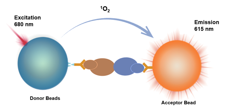
Homogeneous Immuno Chemiluminescence Assay (HICA) is a homogeneous immunoassay method based on energy transfer between donor beads and acceptor beads at close proximity, resulting in luminescence.
Donor beads recognize Protein 1 (Tag1 label), while Acceptor beads recognize Protein 2 (Tag2 label). When Protein 1 binds to Protein 2, the distance between the beads becomes less than 200nm. Upon excitation at 680nm, the donor beads generate singlet oxygen, which diffuses to the acceptor beads. The acceptor beads then undergo a redox reaction, emitting light at 615nm. The signal intensity is directly proportional to the strength of the protein interaction.
This product features a simple operation process, requires no washing, and offers high speed and sensitivity. It is capable of detecting weak interactions.
Components
Specification |
Fill Volume |
250 μg |
50 μL |
5 mg |
1 mL |
25 mg |
1 mL x 5 |
Protocol
# Translation Result:```html
【Required Reagents】
Name |
Catalog Number |
| Anti-Rabbit IgG Acceptor Beads | UA086095 |
| Streptavidin Donor Beads | UA086104 |
| Universal Buffer 1 | UA086113 |
【Detection Protocol for Reference】
Detection Process |
Detection Protocol 1 (37°C Rapid Detection) |
Detection Protocol 2 (Room Temperature Detection) |
Step 1: |
4μL Tag1-M1 +4μL Tag2-M2+ 6μL Acceptor Beads,Light-protected/Green light |
4μL Tag1-M1 +4μL Tag2-M2+ 6μL Acceptor Beads,Light-protected/Green light |
Incubation |
37°C with shaking for 20 minutes,Light-protected/Green light | Room temperature incubation for 60 minutes,Light-protected/Green light |
Step 2: |
Add 6μL Donor Beads,Light-protected/Green light |
Add 6μL Donor Beads,Light-protected/Green light |
Incubation |
37°C with shaking for 10 minutes,Light-protected/Green light |
Room temperature incubation for 30 minutes,Light-protected/Green light |
Reading |
Instrument reading |
Instrument reading |
【Performance Validation】
•Sample Preparation:
Biotinylated rabbit IgG (Bio-rIgG) was pre-diluted to 15μg/mL (100nM) using Universal Buffer 1 as stock solution, then serially diluted according to the following scheme:
ID |
Final Concentration (nM) |
Universal Buffer 1 Volume (μL) |
High Concentration Addition Volume (μL) |
C12 |
1.0E+01 |
210 |
90μL stock solution |
C11 |
3.0E+00 |
210 |
90μL C12 |
|
C10 |
1.0E+00 |
180 |
90μL C11 |
C9 |
3.0E-01 |
210 |
90μL C10 |
C8 |
1.0E-01 |
180 |
90μL C9 |
|
C7极e="border:1px solid #000000;text-align:center;vertical-align:middle;"> 3.0E-02 |
210 |
90μL C8 |
|
C6 |
1.0E-02 |
180 |
90μL C7 |
C5 |
3.0E-03 |
210 |
90μL C6 |
C4 |
1.0E-03 |
180 |
90μL C5 |
C3 |
3.0E-04 |
210 |
90μL C4 |
C2 |
1.0E-04 |
180 |
90μL C3 |
C1 |
0 |
180 |
/ |
•Detection Reagent Preparation:
Name |
Preparation Concentration |
Diluent |
| Anti-Rabbit IgG Acceptor Beads | 25 μg/mL |
Universal Buffer 1 |
| Streptavidin Donor Beads | 25 μg/mL |
Universal Buffer 1 |
•37°C Incubation Mode Results:
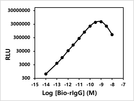
Maximum signal: 4612453
Minimum signal: 646
EC50= 0.123 nM
•Room Temperature Incubation Mode Results:
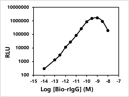
Maximum signal: 1673088
Minimum signal: 305
EC50= 0.111 nM
```Guidelines