Product Specification
| Host |
Rabbit |
| Antigen |
MCM3 |
| Synonyms |
DNA polymerase alpha holoenzyme-associated protein P1,P1-MCM3,RLF subunit beta,p102 |
| Immunogen |
Synthetic Peptide |
| Location |
Nucleus, Chromosome, Intracellular |
| Accession |
P25205 |
| Clone Number |
SDT-048-10 |
| Antibody Type |
Rabbit mAb |
| Application |
WB, IHC-P, ICC, ICFCM, IP |
| Reactivity |
Hu |
| Predicted Reactivity |
Ms, Av, Or, Bv |
| Purification |
Protein A |
| Concentration |
0.5mg/ml |
| Conjugation |
Unconjugated |
| Physical Appearance |
Liquid |
| Storage Buffer |
PBS, 40% Glycerol, 0.05%BSA, 0.03% Proclin 300 |
| Stability & Storage |
12 months from date of receipt / reconstitution, -20 °C as supplied |
Dilution
| application |
dilution |
species |
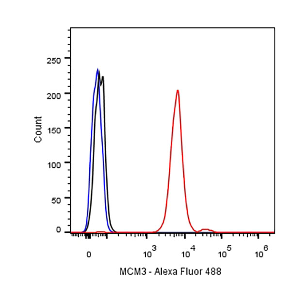
| ICFCM |
1:500 |
|
| ICC |
1:500 |
|
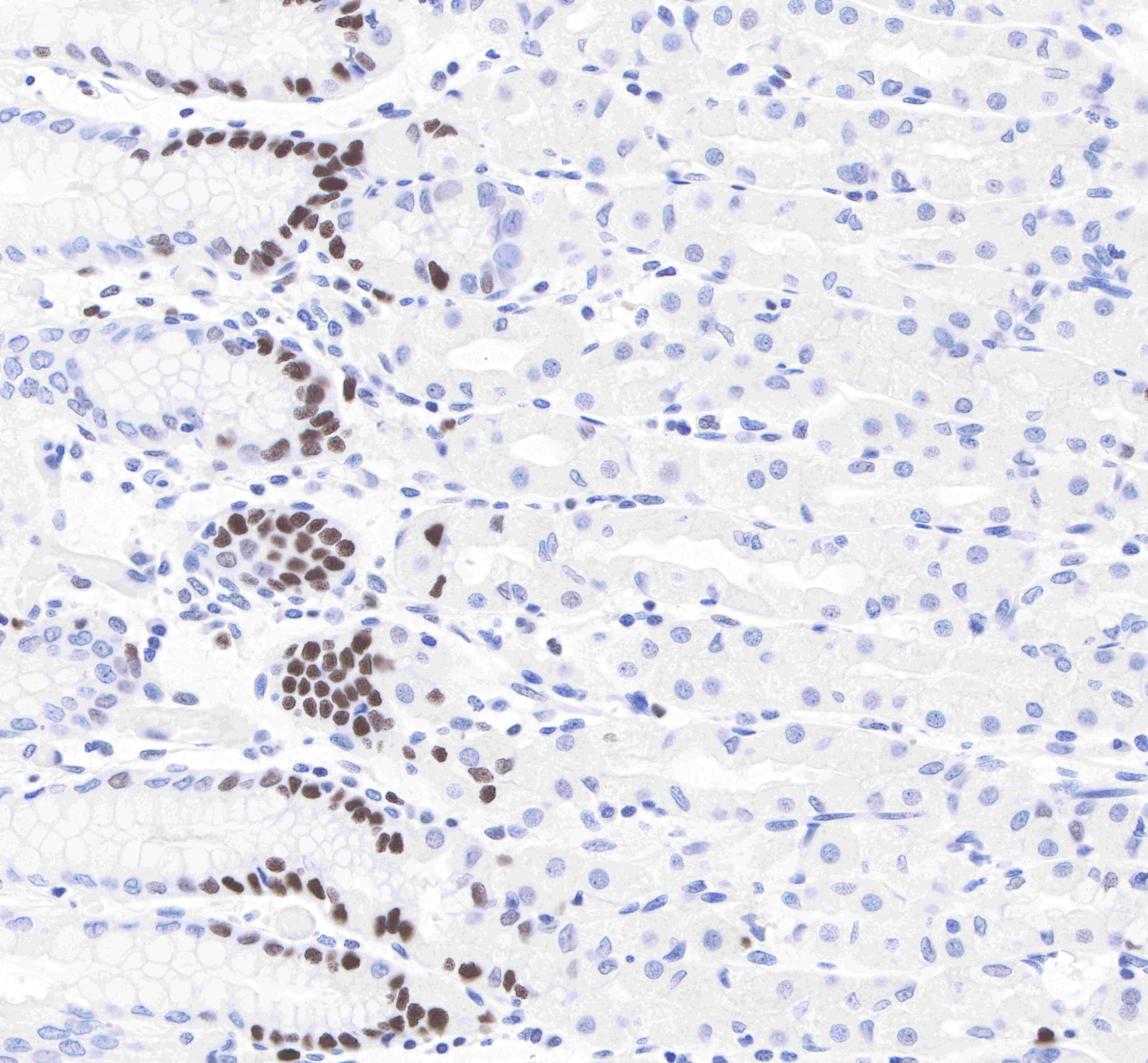
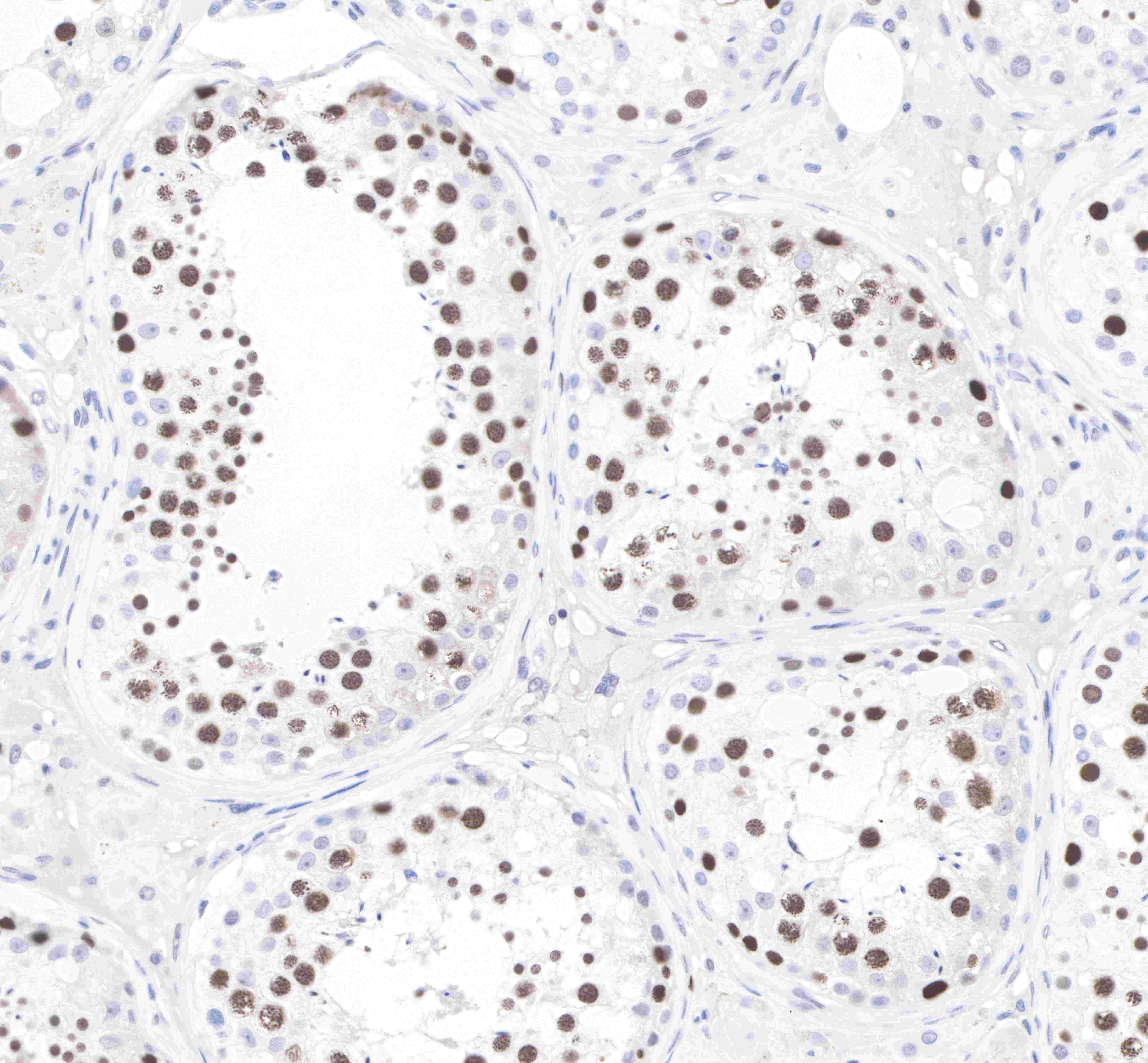
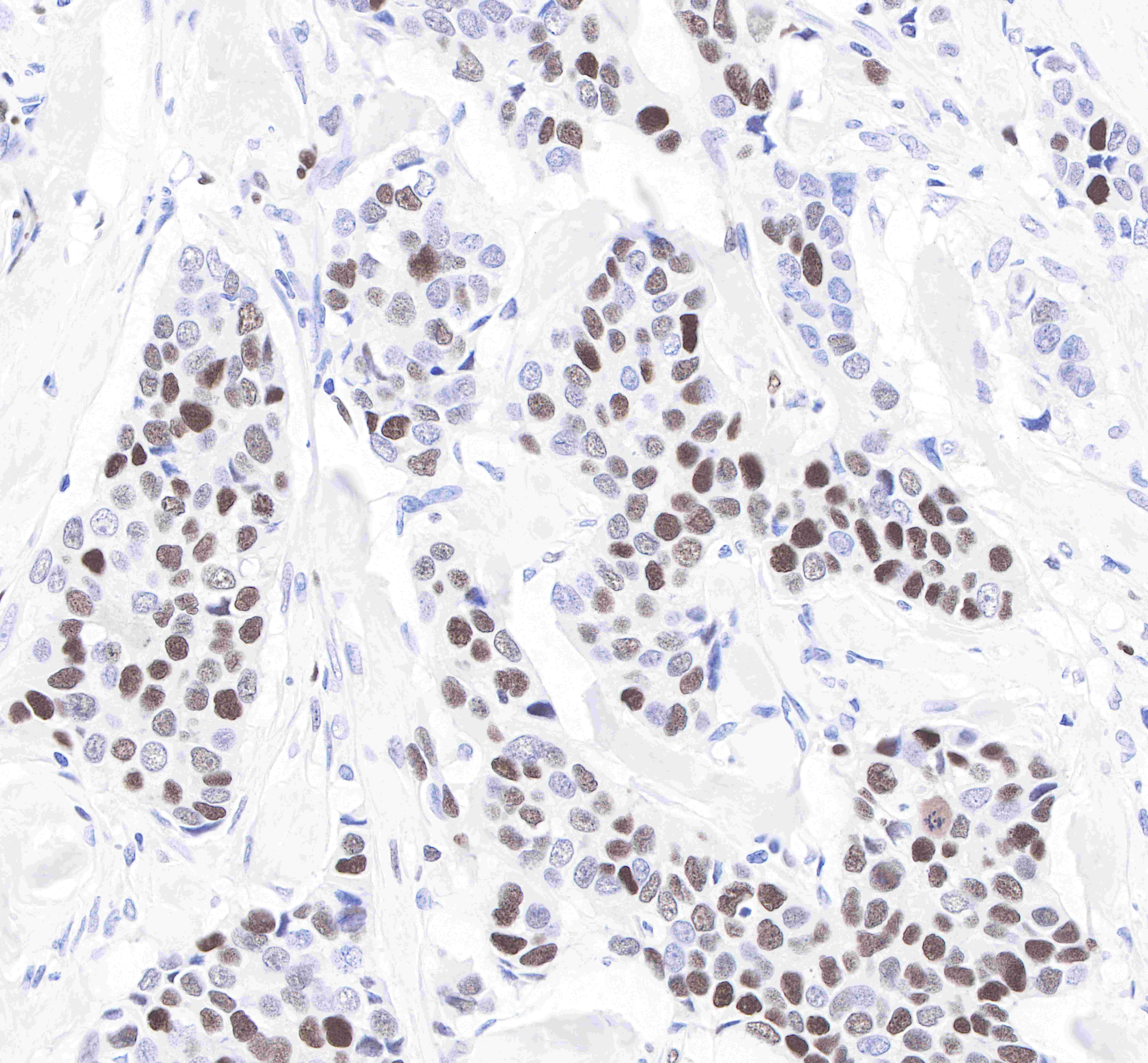
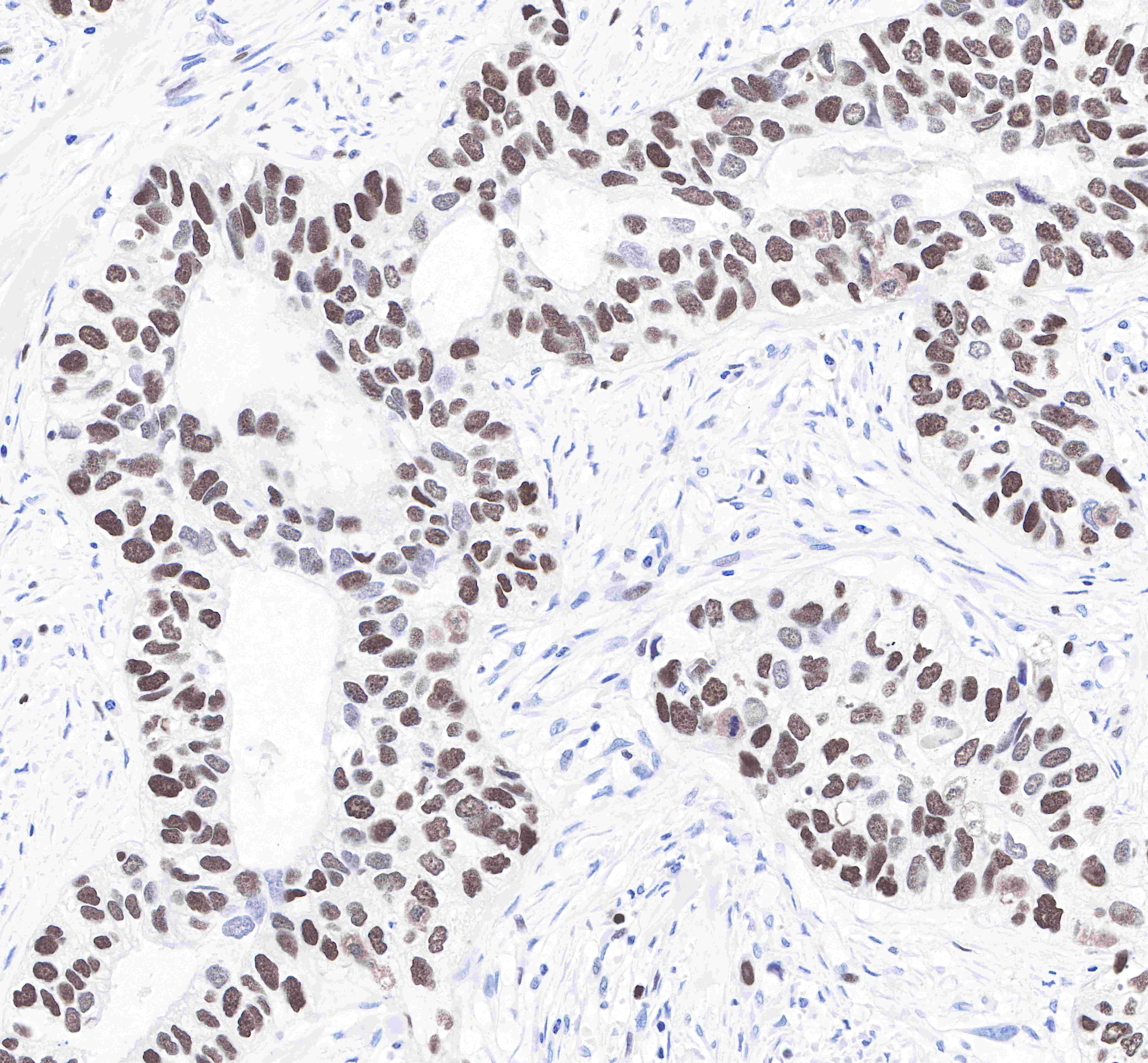
| IHC-P |
1: 2000 |
|
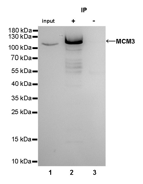
| IP |
1:25 |
|
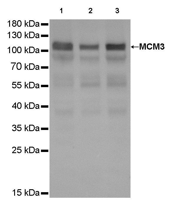
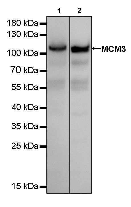
| WB |
1:2000 |
|
Background
DNA replication licensing factor MCM3 is a protein that in humans is encoded by the MCM3 gene. The protein encoded by this gene is one of the highly conserved mini-chromosome maintenance proteins (MCM) that are involved in the initiation of eukaryotic genome replication. The hexameric protein complex formed by MCM proteins is a key component of the pre-replication complex (pre-RC) and may be involved in the formation of replication forks and in the recruitment of other DNA replication related proteins. This protein is a subunit of the protein complex that consists of MCM2-7. It has been shown to interact directly with MCM5/CDC46. This protein also interacts with, and thus is acetylated by MCM3AP, a chromatin-associated acetyltransferase. The acetylation of this protein inhibits the initiation of DNA replication and cell cycle progression.