WB result of VISTA Recombinant Rabbit mAb
Primary antibody: VISTA Recombinant mAb at 1/1000 dilution
Lane 1: MCF-7 whole cell lysate 20 µg
Lane 2: HeLa whole cell lysate 20 µg
Lane 3: DU 145 whole cell lysate 20 µg
Lane 4: Jurkat whole cell lysate 20 µg
Negative control: MCF-7 whole cell lysate
Secondary antibody: Goat Anti-rabbit IgG, (H+L), HRP conjugated at 1/10000 dilution
Predicted MW: 34 kDa
Observed MW: 38~70 kDa
VISTA Recombinant Rabbit mAb (S-1013-5)
VISTA Recombinant Rabbit mAb (S-1013-5)
Price:
Regular price
$100 USD
Regular price
Sale price
$100 USD
Unit price
per
For shipping services or bulk orders, you may request a quotation.
Secure checkout with

View full details
Product Details
Product Details
Product Specification
| Host | Rabbit |
| Antigen | VISTA |
| Synonyms | V-type immunoglobulin domain-containing suppressor of T-cell activation, Platelet receptor Gi24, Stress-induced secreted protein-1 (Sisp-1), V-set domain-containing immunoregulatory receptor, V-set immunoregulatory receptor, VSIR, C10orf54, SISP1 |
| Immunogen | Recombinant Protein |
| Location | Cell membrane |
| Accession | Q9H7M9 |
| Clone Number | S-1013-5 |
| Antibody Type | Recombinant mAb |
| Isotype | IgG |
| Application | WB, IHC-P, IP |
| Reactivity | Hu |
| Purification | Protein A |
| Concentration | 0.5 mg/ml |
| Conjugation | Unconjugated |
| Physical Appearance | Liquid |
| Storage Buffer | PBS, 40% Glycerol, 0.05% BSA, 0.03% Proclin 300 |
| Stability & Storage | 12 months from date of receipt / reconstitution, -20 °C as supplied |
Dilution
| application | dilution | species |
| WB | 1:1000 | null |
| IHC-P | 1:250 | null |
| IP | 1:50 | null |
Background
The VISTA protein, also known as V-domain immunoglobulin suppressor of T cell activation, plays a crucial role in immune regulation. The VISTA protein primarily functions as a T cell inhibitory ligand and receptor, participating in immune regulation by directly suppressing T cell function. As a co-inhibitory ligand, VISTA can act on yet undefined T cell-expressed receptors to inhibit the proliferation of CD4+ and CD8+ T cells, the production of IL-2 and IFN-γ, and promote the conversion of naive T cells into FoxP3+ regulatory T cells. As a co-inhibitory receptor, VISTA can suppress T cell function independently of antigen-presenting cells (APCs). VISTA-deficient mice exhibit spontaneous T cell activation, high levels of proinflammatory cytokine secretion, and more severe experimental autoimmune encephalomyelitis.
Picture
Picture
Western Blot
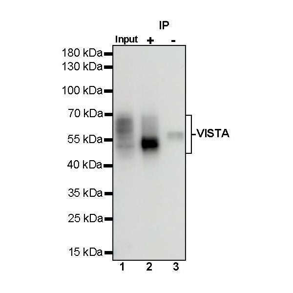
IP
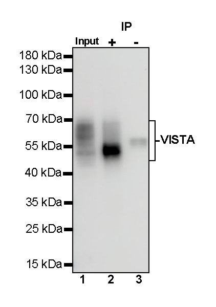
VISTA Rabbit mAb at 1/50 dilution (1 µg) immunoprecipitating VISTA in 0.4 mg DU 145 whole cell lysate.
Western blot was performed on the immunoprecipitate using VISTA Rabbit mAb at 1/1000 dilution.
Secondary antibody (HRP) for IP was used at 1/1000 dilution.
Lane 1: DU 145 whole cell lysate 20 µg (Input)
Lane 2: VISTA Rabbit mAb IP in DU 145 whole cell lysate
Lane 3: Rabbit monoclonal IgG IP in DU 145 whole cell lysate
Predicted MW: 34 kDa
Observed MW: 38~70 kDa
Immunohistochemistry
IHC shows positive staining in paraffin-embedded human cerebral cortex. Anti-VISAT antibody was used at 1/250 dilution, followed by a HRP Polymer for Mouse & Rabbit IgG (ready to use). Counterstained with hematoxylin. Heat mediated antigen retrieval with Tris/EDTA buffer pH9.0 was performed before commencing with IHC staining protocol.
IHC shows positive staining in paraffin-embedded human placenta. Anti-VISAT antibody was used at 1/250 dilution, followed by a HRP Polymer for Mouse & Rabbit IgG (ready to use). Counterstained with hematoxylin. Heat mediated antigen retrieval with Tris/EDTA buffer pH9.0 was performed before commencing with IHC staining protocol.
IHC shows positive staining in paraffin-embedded human cervical squamous cell carcinoma. Anti-VISAT antibody was used at 1/250 dilution, followed by a HRP Polymer for Mouse & Rabbit IgG (ready to use). Counterstained with hematoxylin. Heat mediated antigen retrieval with Tris/EDTA buffer pH9.0 was performed before commencing with IHC staining protocol.
IHC shows positive staining in paraffin-embedded human colon cancer. Anti-VISAT antibody was used at 1/250 dilution, followed by a HRP Polymer for Mouse & Rabbit IgG (ready to use). Counterstained with hematoxylin. Heat mediated antigen retrieval with Tris/EDTA buffer pH9.0 was performed before commencing with IHC staining protocol.
IHC shows positive staining in paraffin-embedded human lung adenocarcinoma. Anti-VISAT antibody was used at 1/250 dilution, followed by a HRP Polymer for Mouse & Rabbit IgG (ready to use). Counterstained with hematoxylin. Heat mediated antigen retrieval with Tris/EDTA buffer pH9.0 was performed before commencing with IHC staining protocol.