WB result of RPE65 Recombinant Rabbit mAb
Primary antibody: RPE65 Recombinant Rabbit mAb at 1/1000 dilution
Lane 1: mouse eye lysate 20 µg
Secondary antibody: Goat Anti-rabbit IgG, (H+L), HRP conjugated at 1/10000 dilution
Predicted MW: 61 kDa
Observed MW: 65 kDa
RPE65 Recombinant Rabbit mAb (S-525-124)
RPE65 Recombinant Rabbit mAb (S-525-124)
Price:
Regular price
$130.00 SGD
Regular price
Sale price
$130.00 SGD
Unit price
per
For shipping services or bulk orders, you may request a quotation.
Secure checkout with

View full details
Product Details
Product Details
Product Specification
| Host | Rabbit |
| Antigen | RPE65 |
| Synonyms | Retinoid isomerohydrolase, All-trans-retinyl-palmitate hydrolase, Lutein isomerase, Meso-zeaxanthin isomerase, Retinal pigment epithelium-specific 65 kDa protein, Retinol isomerase |
| Immunogen | Synthetic Peptide |
| Location | Cytoplasm, Cell membrane |
| Accession | Q16518 |
| Clone Number | S-525-124 |
| Antibody Type | Recombinant mAb |
| Isotype | IgG |
| Application | WB, IHC-P, IP, IF |
| Reactivity | Hu, Ms, Rt |
| Predicted Reactivity | Dg, Pr |
| Purification | Protein A |
| Concentration | 0.5 mg/ml |
| Conjugation | Unconjugated |
| Physical Appearance | Liquid |
| Storage Buffer | PBS, 40% Glycerol, 0.05% BSA, 0.03% Proclin 300 |
| Stability & Storage | 12 months from date of receipt / reconstitution, -20 °C as supplied |
Dilution
| application | dilution | species |
| WB | 1:1000 | |
| IP | 1:50 | |
| IHC-P | 1:1000 | |
| IF | 1:1000 |
Background
The RPE65 protein, also known as Retinoid isomerohydrolase, is a crucial protein produced in the retinal pigment epithelium (RPE) cells. PE65 protein serves as a key isomerohydrolase in the visual cycle, involved in the regeneration of 11-cis-retinal, which is the chromophore of rod and cone cell visual pigments. It catalyzes the cleavage and isomerization of all-trans-retinyl fatty acid esters to produce 11-cis-retinol, which is further oxidized by 11-cis-retinol dehydrogenase to 11-cis-retinal. RPE65 protein exists in two forms: soluble sRPE65 and membrane-bound mRPE65. The latter is palmitoylated and serves as a palmitoyl donor for lecithin retinol acyltransferase (LRAT), which catalyzes a step in the regeneration of vitamin A to all-trans-retinol.
Picture
Picture
Western Blot
WB result of RPE65 Recombinant Rabbit mAb
Primary antibody: RPE65 Recombinant Rabbit mAb at 1/1000 dilution
Lane 1: rat eye lysate 20 µg
Secondary antibody: Goat Anti-rabbit IgG, (H+L), HRP conjugated at 1/10000 dilution
Predicted MW: 61 kDa
Observed MW: 65 kDa
IP
RPE65 Rabbit mAb at 1/50 dilution (1 µg) immunoprecipitating RPE65 in 0.4 mg mouse eye lysate.
Western blot was performed on the immunoprecipitate using RPE65 Rabbit mAb at 1/1000 dilution.
Secondary antibody (HRP) for IP was used at 1/1000 dilution.
Lane 1: mouse eye lysate 20 µg (Input)
Lane 2: RPE65 Rabbit mAb IP in mouse eye lysate
Lane 3: Rabbit monoclonal IgG IP in mouse eye lysate
Predicted MW: 61 kDa
Observed MW: 65 kDa
Immunohistochemistry
IHC shows positive staining in paraffin-embedded mouse retina. Anti-RPE65 antibody was used at 1/1000 dilution, followed by a HRP Polymer for Mouse & Rabbit IgG (ready to use). Counterstained with hematoxylin. Heat mediated antigen retrieval with Tris/EDTA buffer pH9.0 was performed before commencing with IHC staining protocol.
IHC shows positive staining in paraffin-embedded rat retina. Anti-RPE65 antibody was used at 1/1000 dilution, followed by a HRP Polymer for Mouse & Rabbit IgG (ready to use). Counterstained with hematoxylin. Heat mediated antigen retrieval with Tris/EDTA buffer pH9.0 was performed before commencing with IHC staining protocol.
Immunofluorescence

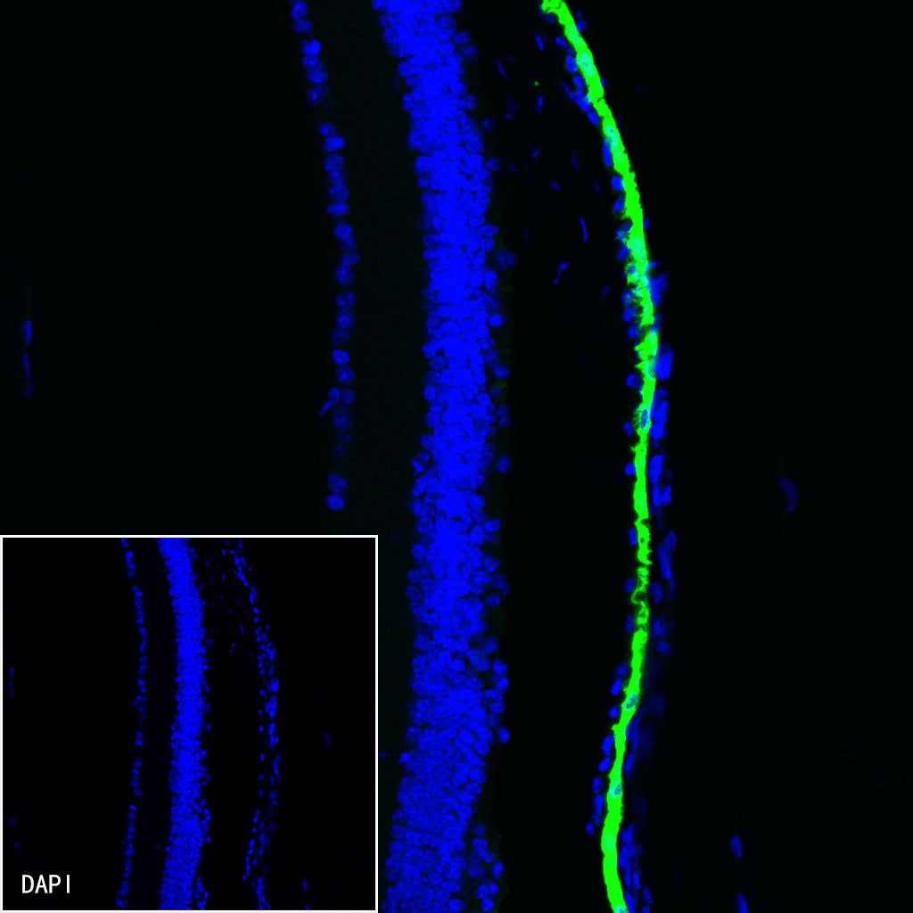
IF shows positive staining in paraffin-embedded mouse retina. Anti-RPE65 antibody was used at 1/1000 dilution (Green) and incubated overnight at 4°C. Goat polyclonal Antibody to Rabbit IgG - H&L (Alexa Fluor® 488) was used as secondary antibody at 1/1000 dilution. Counterstained with DAPI (Blue). Heat mediated antigen retrieval with EDTA buffer pH9.0 was performed before commencing with IF staining protocol.

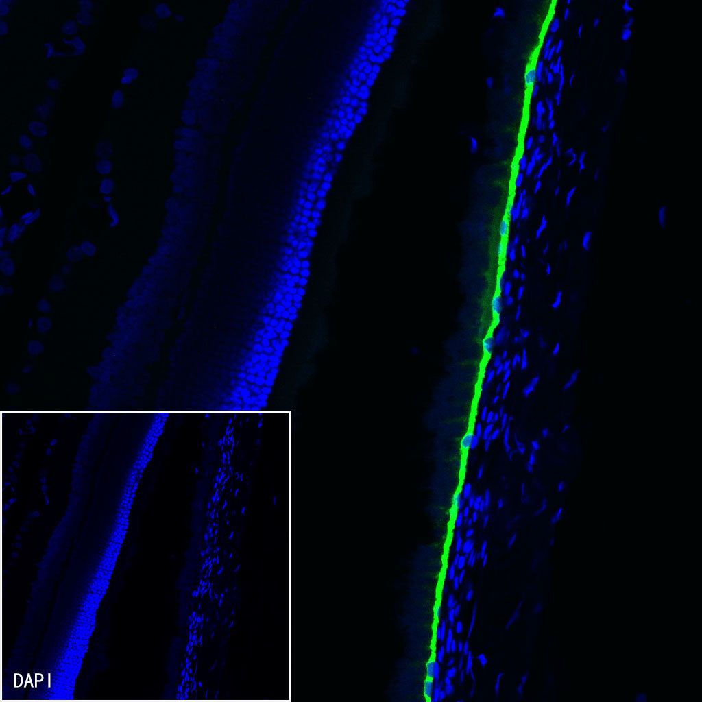
IF shows positive staining in paraffin-embedded rat retina. Anti-RPE65 antibody was used at 1/1000 dilution (Green) and incubated overnight at 4°C. Goat polyclonal Antibody to Rabbit IgG - H&L (Alexa Fluor® 488) was used as secondary antibody at 1/1000 dilution. Counterstained with DAPI (Blue). Heat mediated antigen retrieval with EDTA buffer pH9.0 was performed before commencing with IF staining protocol.