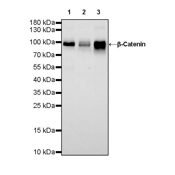
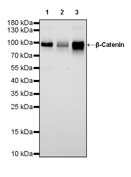
WB result of β-Catenin Rabbit mAb
Primary antibody: β-Catenin Rabbit mAb at 1/1000 dilution
Lane 1: HeLa whole cell lysate 20 µg
Lane 2: HepG2 whole cell lysate 20 µg
Lane 3: A431 whole cell lysate 20 µg
Secondary antibody: Goat Anti-Rabbit IgG, (H+L), HRP conjugated at 1/10000 dilution
Predicted MW: 85kDa
Observed MW: 92kDa
β-Catenin Recombinant Rabbit mAb (SDT-336-23)
β-Catenin Recombinant Rabbit mAb (SDT-336-23)
Price:
Regular price
$100 USD
Regular price
Sale price
$100 USD
Unit price
per
For shipping services or bulk orders, you may request a quotation.
Secure checkout with

View full details
Product Details
Product Details
Product Specification
| Host | Rabbit |
| Antigen | β-Catenin |
| Synonyms | Catenin beta-1, Beta-catenin,CTNNB1 |
| Immunogen | Synthetic Peptide |
| Location | Cytoplasm, Cell membrane |
| Accession | P35222 |
| Clone Number | SDT-336-23 |
| Antibody Type | Recombinant mAb |
| Application | WB, IHC-P, IP, IF |
| Reactivity | Hu, Ms, Rt |
| Purification | Protein A |
| Concentration | 0.5 mg/ml |
| Conjugation | Unconjugated |
| Physical Appearance | Liquid |
| Storage Buffer | PBS, 40% Glycerol, 0.05% BSA, 0.03% Proclin 300 |
| Stability & Storage | 12 months from date of receipt / reconstitution, -20 °C as supplied |
Dilution
| application | dilution | species |
| WB | 1:1000 | |
| IHC-P | 1:500 | |
| IP | 1:50 | |
| IF | 1:500 |
Background
β-catenin is a multifunction protein with a central role in physiological homeostasis. Its abnormal expression leads to various diseases including cancer. In normal physiology, β-catenin either maintains integrity of epithelial tissues or controls transcription of various genes on extracellular instigations. In epithelial tissues, β-catenin functions as a component of the cadherin protein complex and regulates epithelial cell growth and intracellular adhesion [PMID: 23490077].
Picture
Picture
Western Blot
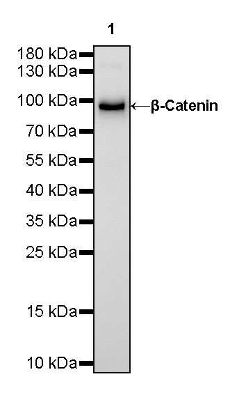
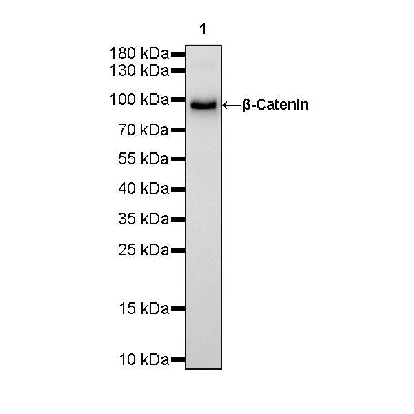
WB result of β-Catenin Rabbit mAb
Primary antibody: β-Catenin Rabbit mAb at 1/1000 dilution
Lane 1: NIH/3T3 whole cell lysate 20 µg
Secondary antibody: Goat Anti-Rabbit IgG, (H+L), HRP conjugated at 1/10000 dilution
Predicted MW: 85kDa
Observed MW: 92kDa
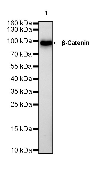
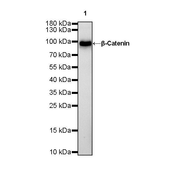
WB result of β-Catenin Rabbit mAb
Primary antibody: β-Catenin Rabbit mAb at 1/1000 dilution
Lane 1: C6 whole cell lysate 20 µg
Secondary antibody: Goat Anti-Rabbit IgG, (H+L), HRP conjugated at 1/10000 dilution
Predicted MW: 85kDa
Observed MW: 92kDa
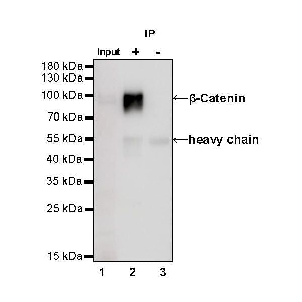
IP
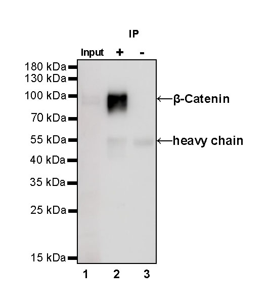
β-Catenin Rabbit mAb at 1/50 dilution (1 µg) immunoprecipitating β-Catenin in 0.4 mg A431 whole cell lysate.
Western blot was performed on the immunoprecipitate using β-Catenin Rabbit mAb at 1/1000 dilution.
Secondary antibody (HRP) for IP was used at 1/400 dilution.
Lane 1: A431 whole cell lysate 20 µg (Input)
Lane 2: β-Catenin Rabbit mAb IP in A431 whole cell lysate
Lane 3: Rabbit monoclonal IgG IP in A431 whole cell lysate
Predicted MW: 85 kDa
Observed MW: 92 kDa
Immunohistochemistry
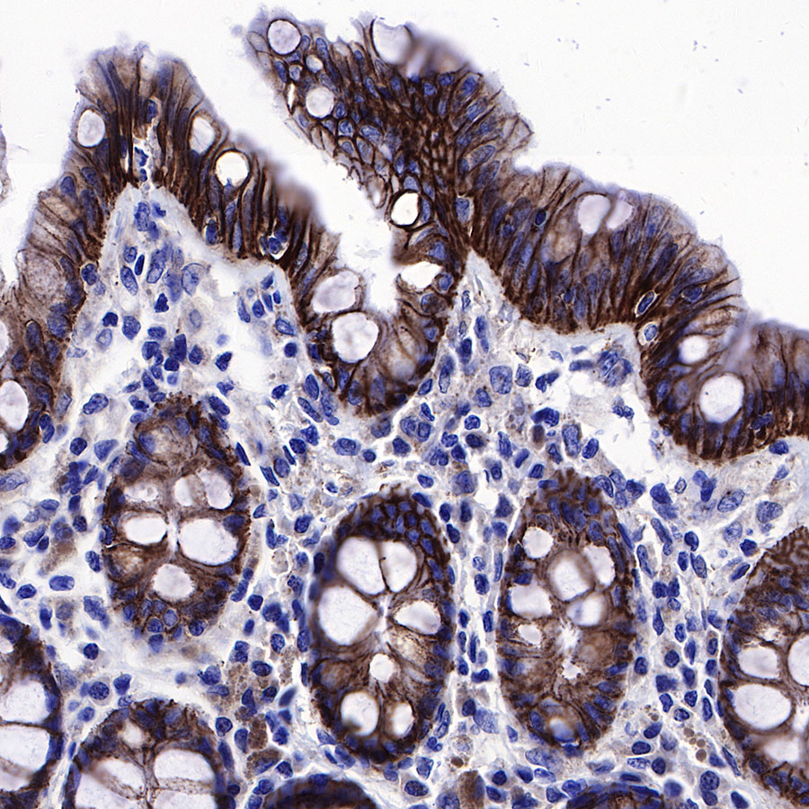
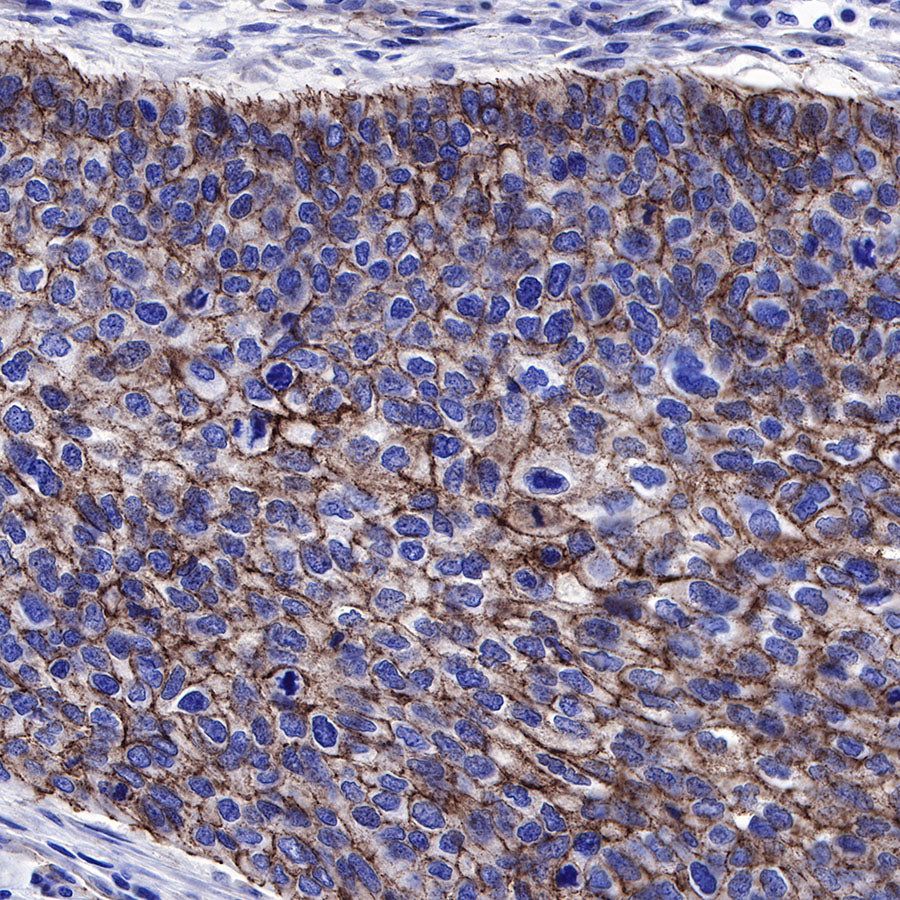
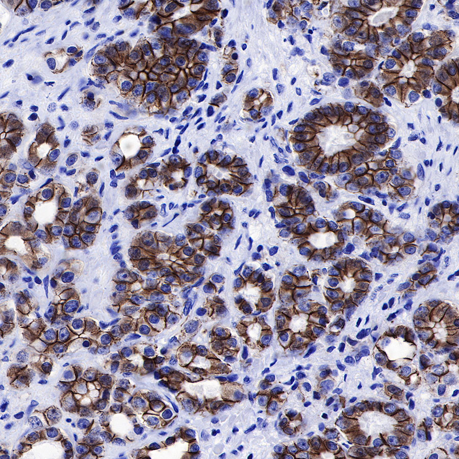
IHC shows positive staining in paraffin-embedded human colon. Anti-β-Catenin antibody was used at 1/500 dilution, followed by a HRP Polymer for Mouse & Rabbit IgG (ready to use). Counterstained with hematoxylin. Heat mediated antigen retrieval with Tris/EDTA buffer pH9.0 was performed before commencing with IHC staining protocol.

IHC shows positive staining in paraffin-embedded human cardiac muscle. Anti-β-Catenin antibody was used at 1/500 dilution, followed by a HRP Polymer for Mouse & Rabbit IgG (ready to use). Counterstained with hematoxylin. Heat mediated antigen retrieval with Tris/EDTA buffer pH9.0 was performed before commencing with IHC staining protocol.
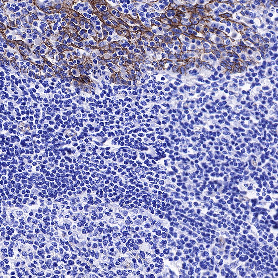
IHC shows positive staining in paraffin-embedded human tonsil. Anti-β-Catenin antibody was used at 1/500 dilution, followed by a HRP Polymer for Mouse & Rabbit IgG (ready to use). Counterstained with hematoxylin. Heat mediated antigen retrieval with Tris/EDTA buffer pH9.0 was performed before commencing with IHC staining protocol.
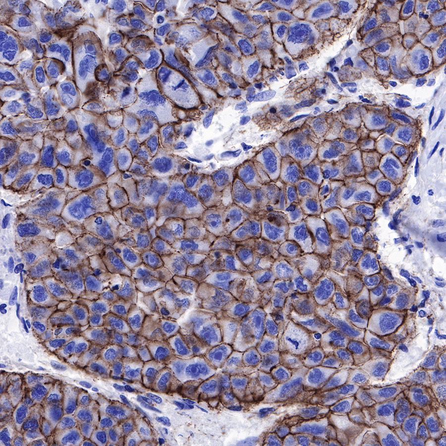
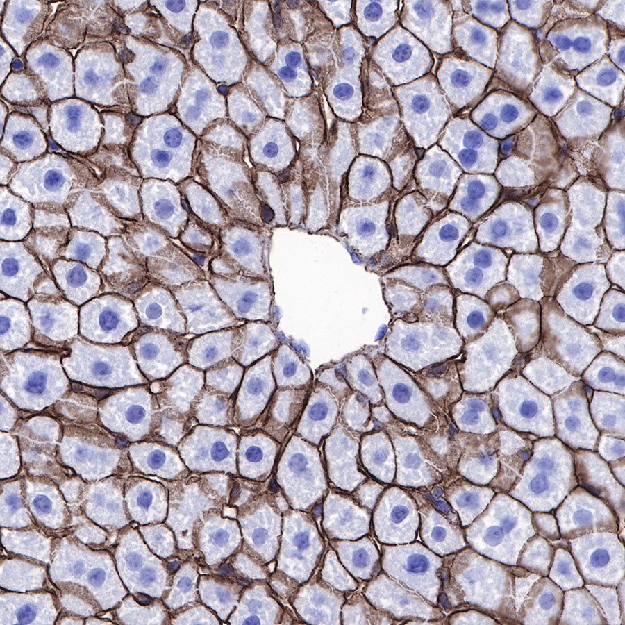
IHC shows positive staining in paraffin-embedded human hepatocellular carcinoma. Anti-β-Catenin antibody was used at 1/500 dilution, followed by a HRP Polymer for Mouse & Rabbit IgG (ready to use). Counterstained with hematoxylin. Heat mediated antigen retrieval with Tris/EDTA buffer pH9.0 was performed before commencing with IHC staining protocol.
IHC shows positive staining in paraffin-embedded human lung adenocarcinoma. Anti-β-Catenin antibody was used at 1/500 dilution, followed by a HRP Polymer for Mouse & Rabbit IgG (ready to use). Counterstained with hematoxylin. Heat mediated antigen retrieval with Tris/EDTA buffer pH9.0 was performed before commencing with IHC staining protocol.

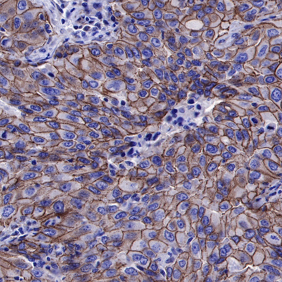
IHC shows positive staining in paraffin-embedded human lung squamous cell carcinoma. Anti-β-Catenin antibody was used at 1/500 dilution, followed by a HRP Polymer for Mouse & Rabbit IgG (ready to use). Counterstained with hematoxylin. Heat mediated antigen retrieval with Tris/EDTA buffer pH9.0 was performed before commencing with IHC staining protocol.
IHC shows positive staining in paraffin-embedded human prostatic carcinoma. Anti-β-Catenin antibody was used at 1/500 dilution, followed by a HRP Polymer for Mouse & Rabbit IgG (ready to use). Counterstained with hematoxylin. Heat mediated antigen retrieval with Tris/EDTA buffer pH9.0 was performed before commencing with IHC staining protocol.

IHC shows positive staining in paraffin-embedded mouse liver. Anti-β-Catenin antibody was used at 1/500 dilution, followed by a HRP Polymer for Mouse & Rabbit IgG (ready to use). Counterstained with hematoxylin. Heat mediated antigen retrieval with Tris/EDTA buffer pH9.0 was performed before commencing with IHC staining protocol.

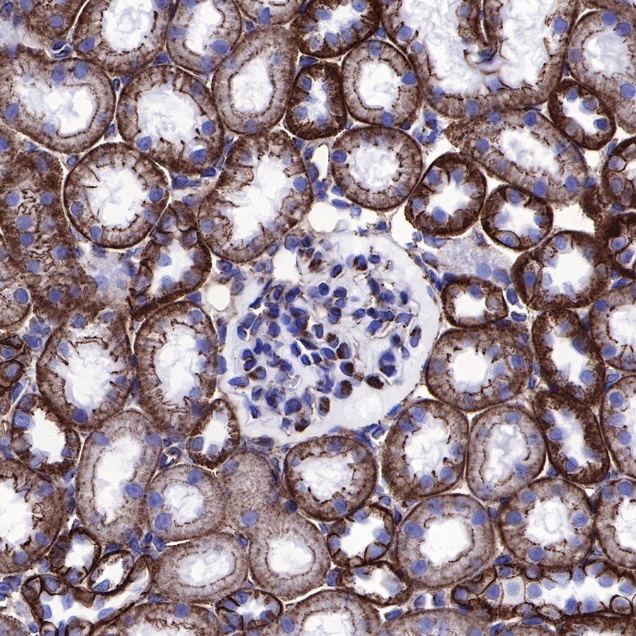
IHC shows positive staining in paraffin-embedded rat kidney. Anti-β-Catenin antibody was used at 1/500 dilution, followed by a HRP Polymer for Mouse & Rabbit IgG (ready to use). Counterstained with hematoxylin. Heat mediated antigen retrieval with Tris/EDTA buffer pH9.0 was performed before commencing with IHC staining protocol.
Immunofluorescence
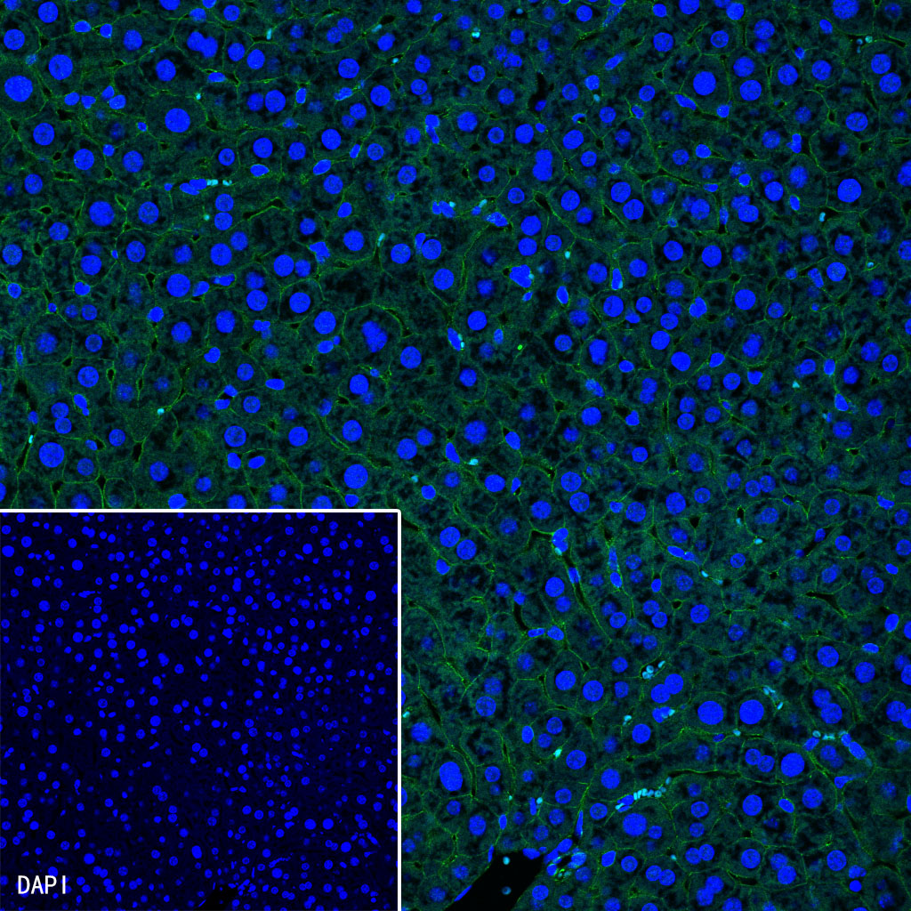
IF shows positive staining in paraffin-embedded mouse liver. Anti-β-Catenin antibody was used at 1/500 dilution (Green) and incubated overnight at 4°C. Goat polyclonal Antibody to Rabbit IgG - H&L (Alexa Fluor® 488) was used as secondary antibody at 1/1000 dilution. Counterstained with DAPI (Blue). Heat mediated antigen retrieval with Tris/EDTA buffer pH9.0 was performed before commencing with IF staining protocol.
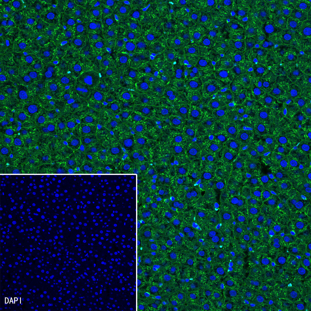
IF shows positive staining in paraffin-embedded rat liver. Anti-β-Catenin antibody was used at 1/500 dilution (Green) and incubated overnight at 4°C. Goat polyclonal Antibody to Rabbit IgG - H&L (Alexa Fluor® 488) was used as secondary antibody at 1/1000 dilution. Counterstained with DAPI (Blue). Heat mediated antigen retrieval with Tris/EDTA buffer pH9.0 was performed before commencing with IF staining protocol.