β-actin Recombinant Rabbit mAb (SDT-R156)
β-actin Recombinant Rabbit mAb (SDT-R156)
Price:
Regular price
$70 USD
Regular price
Sale price
$70 USD
Unit price
per
For shipping services or bulk orders, you may request a quotation.
Secure checkout with

View full details
Product Details
Product Details
Product Specification
| Host | Rabbit |
| Antigen | β-Actin |
| Synonyms | ACTB |
| Immunogen | N/A |
| Location | Cytoplasm, Cytoskeleton |
| Accession | P60709 |
| Clone Number | SDT-R156 |
| Antibody Type | Recombinant mAb |
| Application | WB, IHC-P, ICC, ICFCM |
| Reactivity | Hu, Ms |
| Purification | Protein A |
| Concentration | 0.5 mg/ml |
| Physical Appearance | Liquid |
| Storage Buffer | PBS, 40% Glycerol, 0.05% BSA, 0.03% Proclin 300 |
| Stability & Storage | 12 months from date of receipt / reconstitution, -20 °C as supplied. |
Dilution
| application | dilution | species |
| WB | 1:1000-1:5000 | |
| IHC-P | 1:2000 | |
| ICC | 1:500 | |
| ICFCM | 1:500 |
Background
Beta-actin (human gene and protein abbreviation ACTB/ACTB) is one of six different actin isoforms which have been identified in humans. This is one of the two non-muscle cytoskeletal actins. Actins are highly conserved proteins that are involved in cell motility, structure and integrity. Beta actin is often used in Western blotting as a loading control, to normalize total protein amounts and check for eventual protein degradation in the samples. Its transcript is also commonly used as a housekeeping gene standard in qPCR. Its molecular weight is approximately 42 kDa.
Picture
Picture
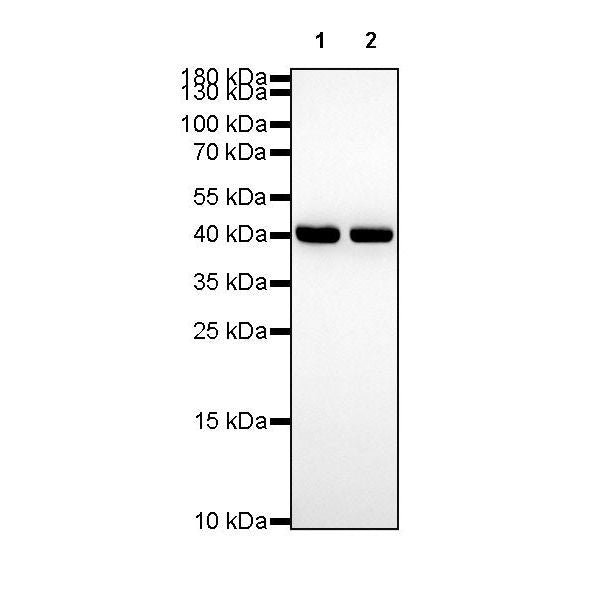
Western Blot
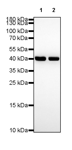
WB result of β-actin Rabbit mAb Primary antibody: β-actin Rabbit mAb at 1/1000 dilution Lane 1: HeLa whole cell lysate 20 µg Lane 2: A549 whole cell lysate 20 µg Secondary antibody: Goat Anti-Rabbit IgG, (H+L), HRP conjugated at 1/10000 dilution Predicted MW: 42kDa Observed MW: 42kDa
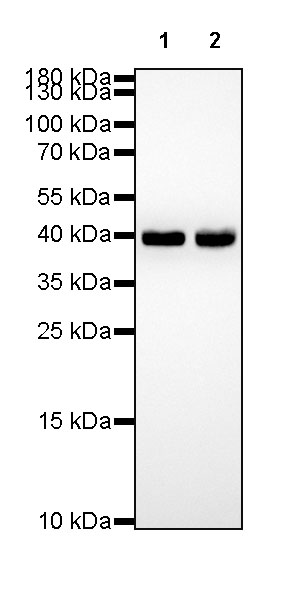
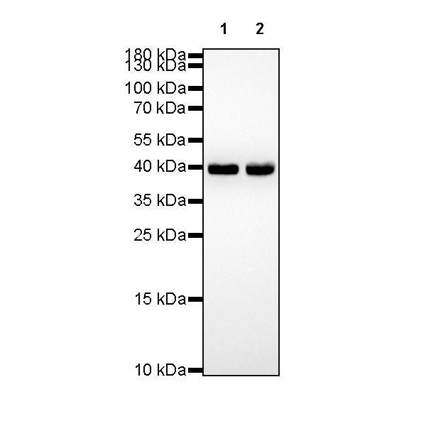
WB result of β-actin Rabbit mAb Primary antibody: β-actin Rabbit mAb at 1/1000 dilution Lane 1: NIH/3T3 whole cell lysate 20 µg Lane 2: mouse kidney lysate 20 µg Secondary antibody: Goat Anti-Rabbit IgG, (H+L), HRP conjugated at 1/10000 dilution Predicted MW: 42kDa Observed MW: 42kDa
FC
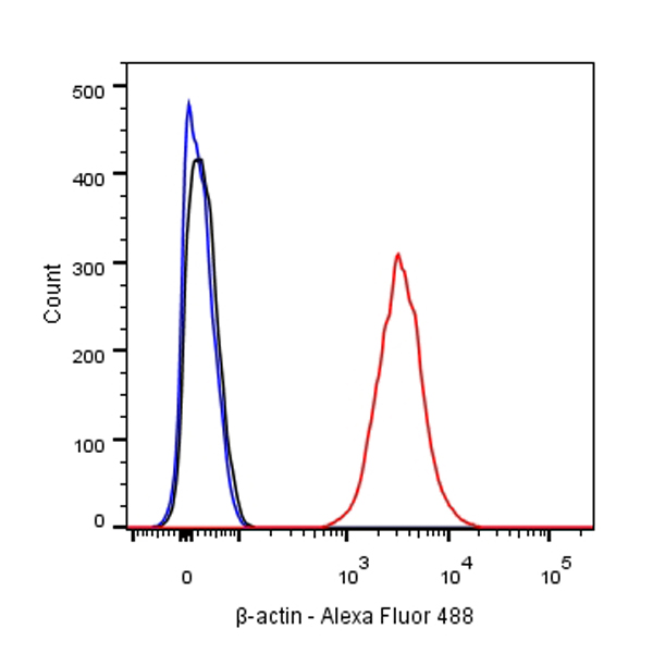
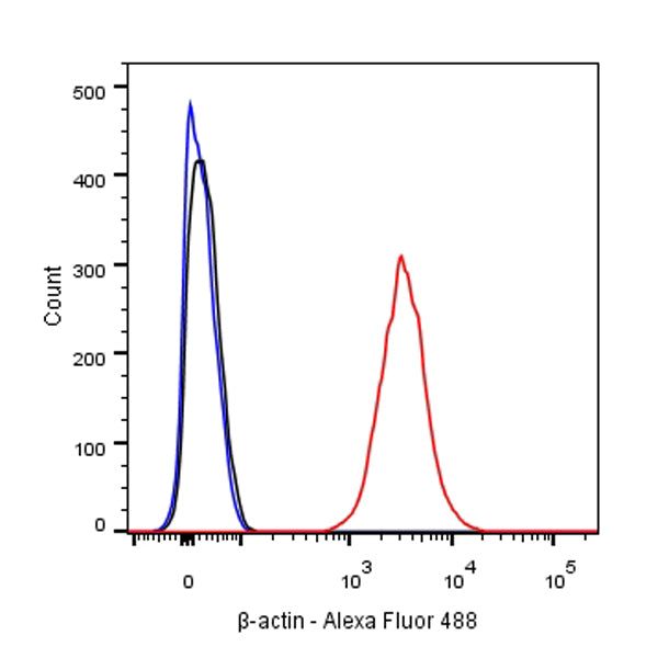
Flow cytometric analysis of HeLa cells labelling β-actin antibody at 1/500 (0.1 μg) dilution/ (red) compared with a Rabbit monoclonal IgG (Black) isotype control and an unlabelled control (cells without incubation with primary antibody and secondary antibody) (Blue). Goat Anti-Rabbit IgG Alexa Fluor® 488 was used as the secondary antibody.
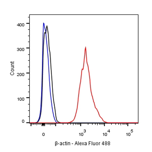
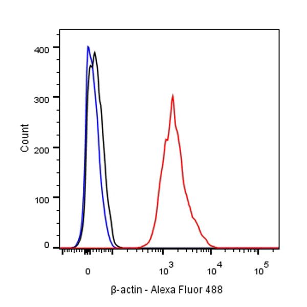
Flow cytometric analysis of NIH/3T3 cells labelling β-actin antibody at 1/500 (0.1 μg) dilution/ (red) compared with a Rabbit monoclonal IgG (Black) isotype control and an unlabelled control (cells without incubation with primary antibody and secondary antibody) (Blue). Goat Anti-Rabbit IgG Alexa Fluor® 488 was used as the secondary antibody.
Immunohistochemistry
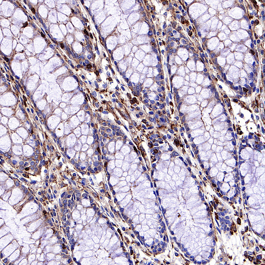
IHC shows positive staining in paraffin-embedded human colon. Anti-β-actin antibody was used at 1/2000 dilution, followed by a HRP Polymer for Mouse & Rabbit IgG (ready to use). Counterstained with hematoxylin. Heat mediated antigen retrieval with Tris/EDTA buffer pH9.0 was performed before commencing with IHC staining protocol.
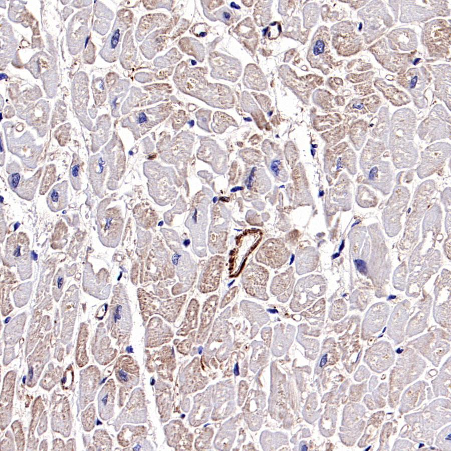
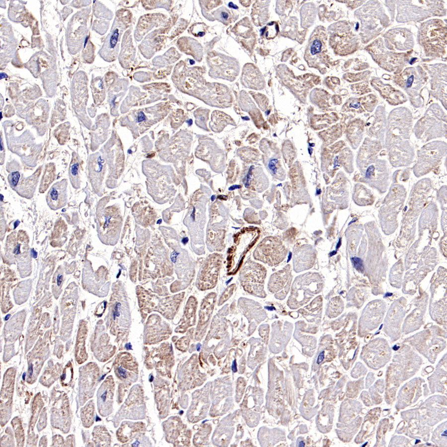
IHC shows positive staining in paraffin-embedded human cardiac muscle. Anti-β-actin antibody was used at 1/2000 dilution, followed by a HRP Polymer for Mouse & Rabbit IgG (ready to use). Counterstained with hematoxylin. Heat mediated antigen retrieval with Tris/EDTA buffer pH9.0 was performed before commencing with IHC staining protocol.
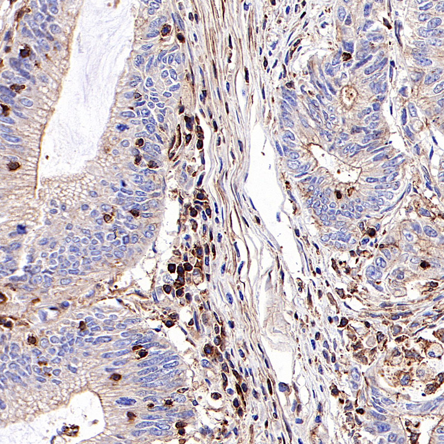
IHC shows positive staining in paraffin-embedded human colon cancer. Anti-β-actin antibody was used at 1/2000 dilution, followed by a HRP Polymer for Mouse & Rabbit IgG (ready to use). Counterstained with hematoxylin. Heat mediated antigen retrieval with Tris/EDTA buffer pH9.0 was performed before commencing with IHC staining protocol.
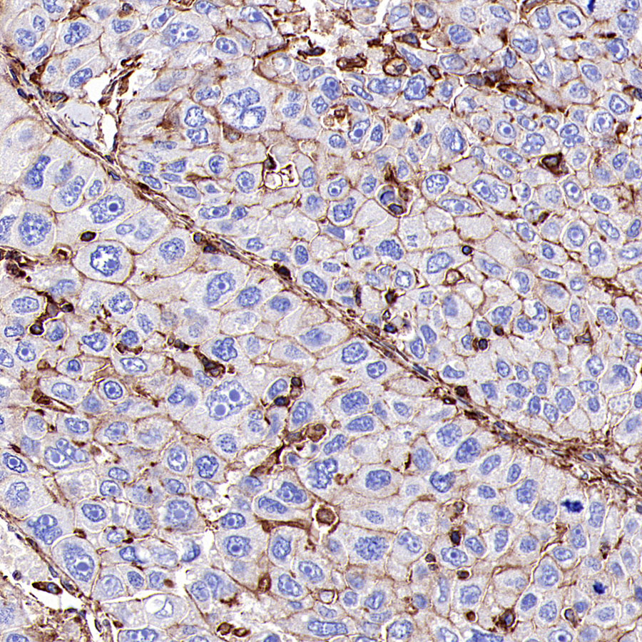
IHC shows positive staining in paraffin-embedded human hepatocellular carcinoma. Anti-β-actin antibody was used at 1/2000 dilution, followed by a HRP Polymer for Mouse & Rabbit IgG (ready to use). Counterstained with hematoxylin. Heat mediated antigen retrieval with Tris/EDTA buffer pH9.0 was performed before commencing with IHC staining protocol.

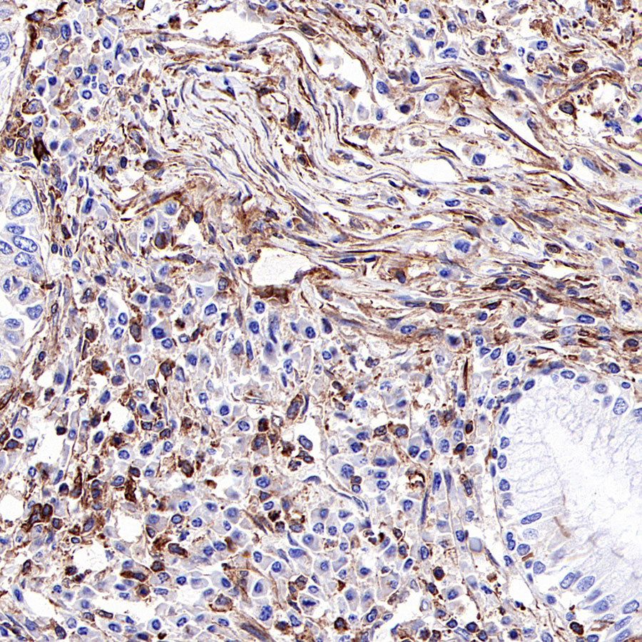
IHC shows positive staining in paraffin-embedded human lung squamous cell carcinoma. Anti-β-actin antibody was used at 1/2000 dilution, followed by a HRP Polymer for Mouse & Rabbit IgG (ready to use). Counterstained with hematoxylin. Heat mediated antigen retrieval with Tris/EDTA buffer pH9.0 was performed before commencing with IHC staining protocol.

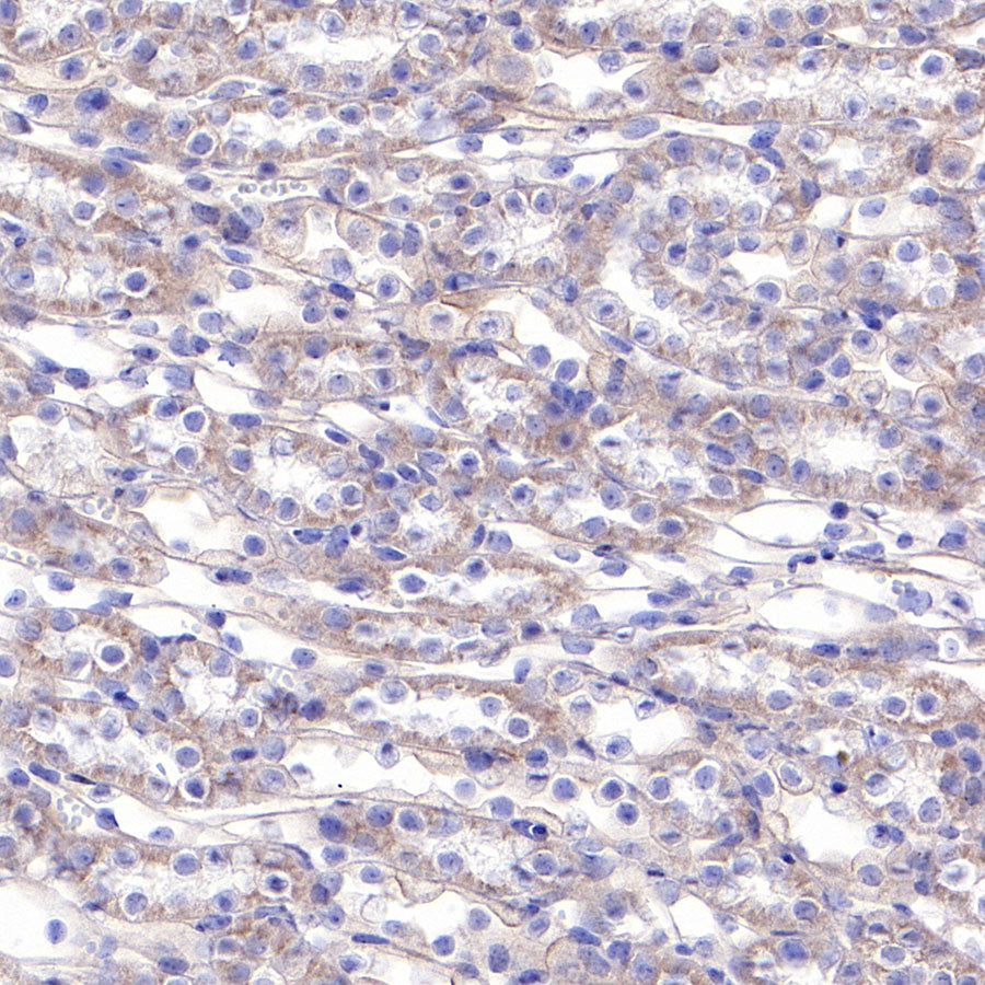
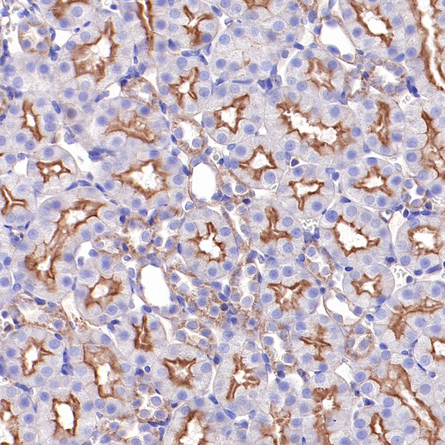
IHC shows positive staining in paraffin-embedded mouse kidney. Anti-β-actin antibody was used at 1/2000 dilution, followed by a HRP Polymer for Mouse & Rabbit IgG (ready to use). Counterstained with hematoxylin. Heat mediated antigen retrieval with Tris/EDTA buffer pH9.0 was performed before commencing with IHC staining protocol.
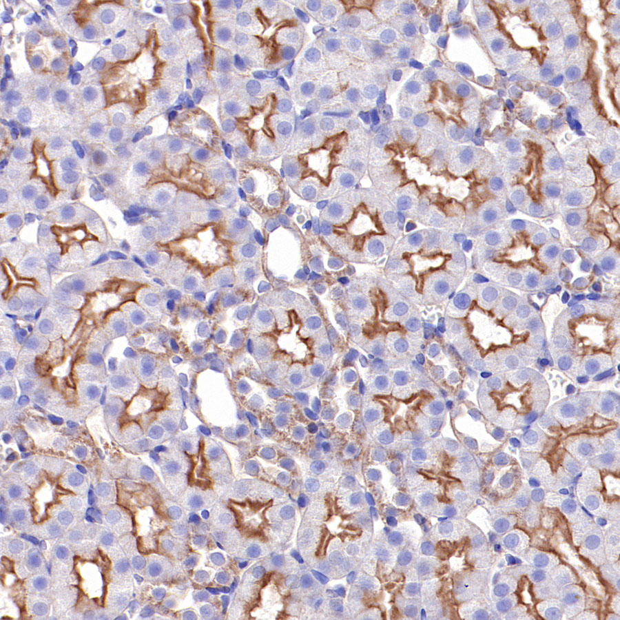
IHC shows positive staining in paraffin-embedded rat kidney. Anti-β-actin antibody was used at 1/2000 dilution, followed by a HRP Polymer for Mouse & Rabbit IgG (ready to use). Counterstained with hematoxylin. Heat mediated antigen retrieval with Tris/EDTA buffer pH9.0 was performed before commencing with IHC staining protocol.
Immunocytochemistry
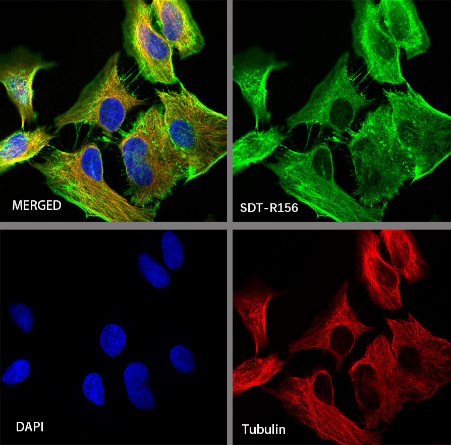
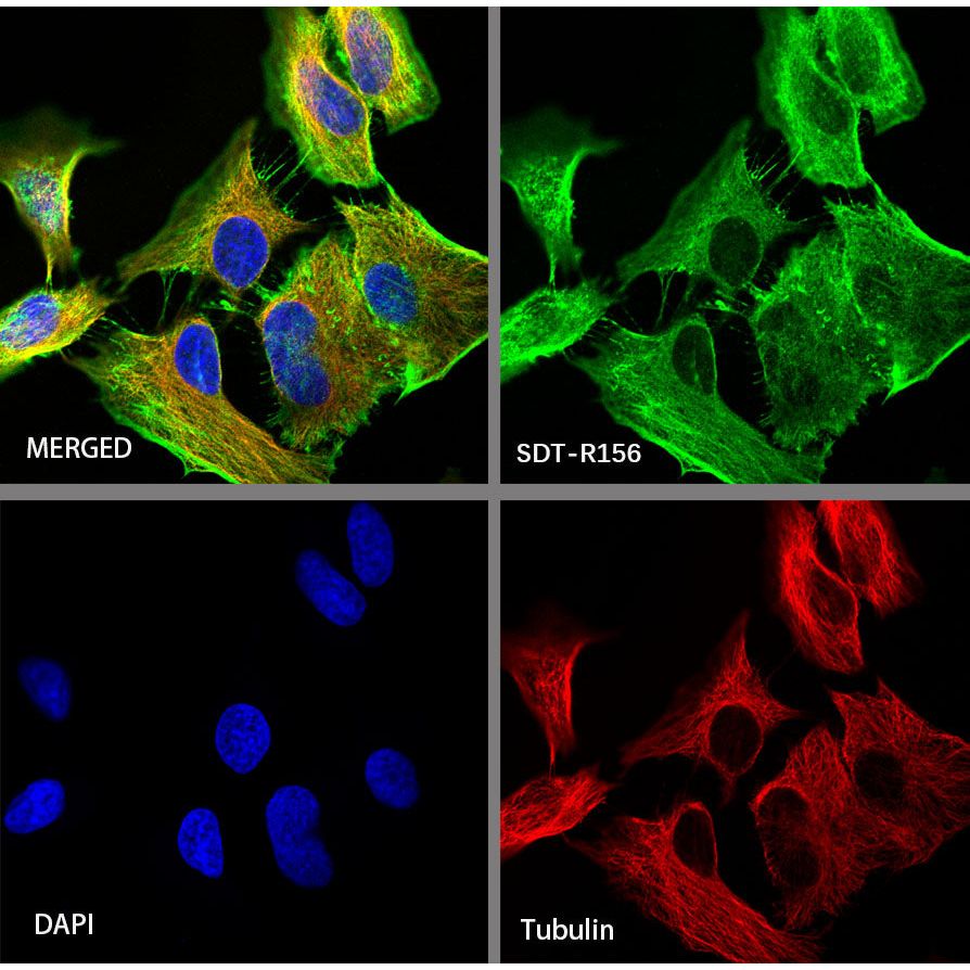
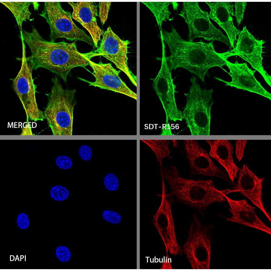
ICC shows positive staining in HeLa cells. Anti-β-actin antibody was used at 1/500 dilution (Green) and incubated overnight at 4°C. Goat polyclonal Antibody to Rabbit IgG - H&L (Alexa Fluor® 488) was used as secondary antibody at 1/1000 dilution. The cells were fixed with 100% ice-cold methanol and permeabilized with 0.1% PBS-Triton X-100. Nuclei were counterstained with DAPI (Blue). Counterstain with tubulin (red).
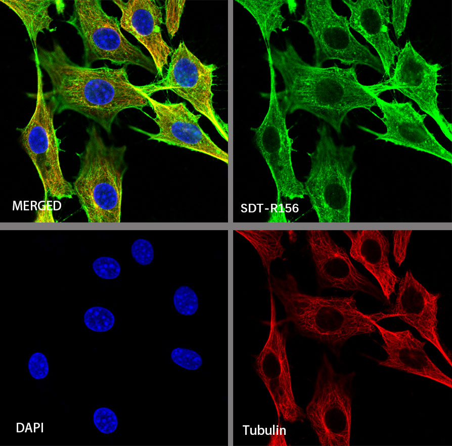
ICC shows positive staining in NIH/3T3 cells. Anti-β-actin antibody was used at 1/500 dilution (Green) and incubated overnight at 4°C. Goat polyclonal Antibody to Rabbit IgG - H&L (Alexa Fluor® 488) was used as secondary antibody at 1/1000 dilution. The cells were fixed with 100% ice-cold methanol and permeabilized with 0.1% PBS-Triton X-100. Nuclei were counterstained with DAPI (Blue). Counterstain with tubulin (red).