UniOne® TR-FRET Human VCB(VHL Complex)Binding Kit
UniOne® TR-FRET Human VCB(VHL Complex)Binding Kit
Product Details
Product Details
Product Specification
| Host | Human |
| Stability & Storage | -80℃ |
Background
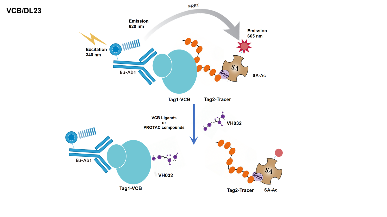
The reagent kit employs homogeneous time-resolved fluorescence technology (TR-FRET) for measuring the interaction between Human VCB (VHL/Elongin-C/Elongin-B Complex) and Human HIF-1α. The Tag2-Tracer is a fluorescently labeled HIF-1α peptide fragment. This method enables simple and rapid high-throughput screening of inhibitors and antibody blockers.
As shown in the figure, the interaction between VCB and Tracer is detected using a Eu-labeled anti-Tag1 antibody (TR-FRET donor) and an Acceptor-labeled SA (TR-FRET acceptor). Since the binding of VCB and Tracer brings the donor antibody and acceptor antibody into proximity, excitation of the donor antibody triggers fluorescence resonance energy transfer (FRET) to the acceptor antibody, resulting in a specific emission signal at 665 nm. VH032 can block the binding of VCB and Tracer, preventing the generation of FRET signals. The stronger the blocking effect of the screened drug on the VCB-Tracer interaction, the lower the signal. The signal is proportional to the degree of interaction between VCB and Tracer. No washing steps are required.
Components
组分 |
浓度 |
100T |
500T |
2500T |
10000T |
储存温度 |
Tag1-VCB protein |
100 × |
5μL |
20μL |
100μL |
400μL |
-80℃ |
Tag2-Tracer |
100 × |
5μL |
20μL |
100μL |
400μL |
-80℃ |
Eu-anti-Tag1 |
50 × |
10μL |
50μL |
250μL |
1000μL |
-80℃ |
SA-Ac |
100 × |
5μL |
25μL |
125μL |
500μL |
-80℃ |
Detection buffer |
1 × |
3mL |
14mL |
70mL |
240mL |
-80℃ |
Protocol
1. Reagent Preparation
1.1 Before use, thaw all reagents at room temperature (room temperature equilibration for at least 30 min). The 384-well shallow well plate reaction system volume is 20 μL (reagent volumes for the reaction system are shown in the table); calculate the required volume before preparation and prepare as needed; the following preparation is for reference only, taking 500 times as an example.
Table 1. Reagent Preparation
Reagent Name |
Preparation |
Volume per Detection Well μL |
Tag1-VCB protein |
Take 20 μL 100 × Tag1-VCB protein stock solution, add to 1.98 mL 1×Detection buffer to dilute to 1×, mix and set aside. | 4μL |
Tag2-Tracer |
Take 20 μL 100 × Tag2-Tracer stock solution, add to 1.98 mL 1×Detection buffer to dilute to 1×, mix and set aside. | 4μL |
|
Detection Reagent Mix
|
Take 50 μL 50× Eu-anti-Tag1 stock solution, add 2.45 mL 1×Detection buffer to dilute to 2.5 mL, mix; Take 25 μL 100× SA-Ac stock solution, add 1×Detection buffer 2.475 mL to dilute to 2.5 mL, mix; Mix both 1:1, this is Detection Mix. |
10μL |
1.2 Gradient Dilution of Test Samples
Taking VH032 as an example, the diluent is 1× Detection buffer. To reduce the interference of matrix effects, it is recommended to use a solution with the same matrix as the test sample for dilution; and adjust the test samples according to actual concentration.
Table 2. Positive Drug VH032 Gradient Dilution (Adjust According to Actual Situation)
|
VH032 Prepared Concentration (nM) |
VH032 Final Concentration (nM) |
Preparation Method |
① |
30,000.0 |
300,000.0 |
2μL 3mM Stock Solution + 18μL 1× Detection buffer |
② |
10000.0 |
100000.0 |
10μL ① +20μL 1× Detection buffer |
③ |
3333.3 |
33333.3 |
10μL ② +20μL 1× Detection buffer |
④ |
1111.1 |
11111.1 |
10μL ③ +20μL 1× Detection buffer |
⑤ |
370.4 |
3703.7 |
10μL ④ +20μL 1× Detection buffer |
⑥ |
123.5 |
1234.6 |
10μL ⑤ +20μL 1× Detection buffer |
⑦ |
41.2 |
411.5 |
10μL ⑥ +20μL 1× Detection buffer |
⑧ |
13.7 |
137.2 |
10μL ⑦ +20μL 1× Detection buffer |
⑨ |
4.6 |
45.7 |
10μL ⑧ +20μL 1× Detection buffer |
Blank |
0.0 |
0 |
20μL 1× Detection buffer |
2. Sample Addition and Controls
2.1 Sample Well (Sample): Add 2 μL test sample (gradient dilution), 4 μL Tag1-VCB protein working solution, 4 μL Tag2-Tracer working solution, 10 μL Detection Reagent Mix sequentially into the 384-well shallow well plate.
2.2 Maximum Signal Control: Add 2 μL Detection buffer, 4 μL Tag1-VCB protein working solution, 4 μL Tag2-Tracer working solution, 10 μL Detection Reagent Mix sequentially into the 384-well shallow well plate.
2.3 Negative Control (Negative control): Add 10 μL Detection buffer, 10 μL Detection Reagent Mix sequentially into the 384-well shallow well plate.
After adding all samples, centrifuge, seal the plate film, and incubate at room temperature for 2 hours.
|
Sample (Sample) |
Maximum Signal Control |
Negative Control (NC) |
Step 1 |
2μL Test Sample |
2μL 1×Detection buffer |
10μL 1×Detection buffer |
4μL Tag1-VCB protein Working Solution | |||
Incubate at Room Temperature 10 min | |||
Step 2 |
4μL Tag2-Tracer Working Solution |
||
10μL Detection Reagent Mix | |||
Seal plate with film, incubate at room temperature for 2h | |||
3.Detection
Detect on a microplate reader compatible with TR-FRET. Excitation light is 320/340 nm, detect emission wavelengths of 620 nm and 665 nm.
[Result Calculation]
1) Calculate Signal Value (Ratio): Fluorescence signal at 665 nm divided by fluorescence signal at 620 nm, then multiplied by 10000.
Ratio = (665/620) ×10000
2) Calculate Net signal based on signal value:
Net signal = (Std-NC)/NC×100
3) Calculate CV (%):
CV (%) = Standard Deviation/Mean Ratio × 100%
[Data Example]
The data below cannot replace data obtained from experiments, it is for demonstration purposes only; results may vary depending on the plate reader instrument.

Note: Recommended Microplate (384-well plate, White, Shallow Well)