UniOne® TR FRET Human PCSK9(D347Y)/LDLR Assay Kit
UniOne® TR FRET Human PCSK9(D347Y)/LDLR Assay Kit
Product Details
Product Details
Product Specification
| Host | Human |
| Stability & Storage | -80℃ |
Background
The kit utilizes homogeneous time-resolved fluorescence (TR-FRET) technology to measure the interaction between Human PCSK9(D347Y) and Human LDLR. PCSK9(D347Y) is a mutant form of PCSK9 with higher affinity for LDLR. This method enables simple, rapid, and high-throughput screening of inhibitors and antibody blockers.
As shown in the figure, the interaction between PCSK9(D347Y) and LDLR is detected using Eu-labeled anti-Tag1 antibody (TR-FRET donor) and Ac-labeled Tag2 antibody (TR-FRET acceptor). The binding of PCSK9(D347Y) to LDLR brings the donor and acceptor antibodies into proximity, allowing the excitation of the donor antibody to trigger fluorescence resonance energy transfer (FRET) to the acceptor antibody, resulting in a specific emission signal at 665 nm. Screened compounds, such as Pep 2-8, block the binding of PCSK9(D347Y) to LDLR, preventing FRET signal generation. The stronger the blocking effect of the screened compound, the lower the signal. The signal is proportional to the degree of interaction between PCSK9(D347Y) and LDLR. No washing steps are required.

Components
Component |
Concentration |
100T |
500T |
2500T |
10000T |
Storage Temperature |
Tag1-LDLR protein |
100× |
5μL |
20μL |
100μL |
400μL |
-80℃ |
Tag2-PCSK9(D347Y) protein |
100× |
5μL |
20μL |
100μL |
400μL |
-80℃ |
Anti-Tag1 Eu antibody |
50× |
10μL |
50μL |
250μL |
1000μL |
-80℃ |
Anti-Tag2 Ac antibody |
12.5× |
40μL |
200μL |
1mL |
4mL |
-80℃ |
Detection buffer |
1× |
2mL |
14mL |
60mL |
240mL |
-80℃ |
Protocol
Here is the translated content in English, preserving the original formatting:```markdown
1. Reagent Preparation
1.1 Thaw all reagents to room temperature before use (allow at least 30 min for equilibration). The reaction system for 384-well plates is 20μL (reagent volumes are shown in the table below). Calculate the required volume before preparation and prepare accordingly. The following preparation is for reference only, using 500 reactions as an example.
Table 1. Reagent Preparation
Reagent Name |
Preparation |
Volume per Well (μL) |
Sample Dilution |
Use 1× Detection Buffer to dilute samples. If the sample stock is in DMSO, maintain consistent DMSO concentration in the system (≤2%). | 2 |
Tag1-LDLR Protein |
Dilute 20μL Tag1-LDLR stock with 1× Detection Buffer to 2mL, mix well. | 4 |
Tag2-PCSK9(D347Y) Protein |
Dilute 20μL Tag2-PCSK9(D347Y) stock with 1× Detection Buffer to 2mL, mix well. | 4 |
Antibody Mix |
Dilute 50μL Anti-Tag1 Eu antibody with 2.45mL 1× Detection Buffer, mix well; dilute 200μL Anti-Tag2 Ac antibody with 2.3mL 1× Detection Buffer, mix well. Combine Anti-Tag1 Eu and Anti-Tag2 Ac antibodies at 1:1 ratio to prepare Antibody Mix. | 10 |
1.2 Sample Gradient Dilution
Using Pep 2-8 as an example, dilute with 1× Detection Buffer. To minimize matrix effects, use the same matrix as the sample. Adjust concentrations as needed.
Table 2. Pep 2-8 Preparation Table (adjust as needed)
Pep 2-8 Prep Conc. (nM) |
Final Conc. (nM) |
Preparation Method |
|
| 1 | 1000000 |
100000 |
12μL 1mM Pep 2-8 |
| 2 | 333333.33 |
33333.33 |
6μL ① +12μL 1× Detection Buffer |
| 3 | 111111.11 |
11111.11 |
5μL ② +10μL 1× Detection Buffer |
| 4 | 37037.04 |
3703.70 |
5μL ③ +10μL 1× Detection Buffer |
| 5 | 12345.68 |
1234.57 |
5μL ④ +10μL 1× Detection Buffer |
| 6 | 4115.23 |
411.52 |
5μL ⑤ +10μL 1× Detection Buffer |
| 7 | 1371.74 |
137.17 |
5μL ⑥ +10μL 1× Detection Buffer |
| 8 | 457.25 |
45.72 |
5μL ⑦ +10μL 1× Detection Buffer |
| 9 | 152.42 |
15.24 |
5μL ⑧ +10μL 1× Detection Buffer |
Blank |
0 |
0 |
10μL 1× Detection Buffer |
2. Sample Loading & Controls
2.1 Test Samples: Add sequentially to 384-well plate—2μL gradient-diluted sample, 4μL Tag2-PCSK9(D347Y), 4μL Tag1-LDLR, 10μL Antibody Mix.
2.2 Blank Control (Blank): Replace sample with 2μL 1× Detection Buffer.
2.3 Eu Control (NC): 10μL 1× Detection Buffer + 10μL Antibody Mix.
After loading, centrifuge, seal, and incubate at RT for 2 hours.
|
Test Sample |
Blank Control |
Eu Control (NC) |
Step 1 |
2μL gradient-diluted sample |
2μL 1× Detection Buffer |
10μL 1× Detection Buffer +10μL Antibody Mix |
4μL Tag2-PCSK9(D347Y) | |||
|
Seal, centrifuge at 1000rpm for 1min, incubate at RT for 10min | |||
Step 2 |
4μL Tag1-LDLR |
||
10μL Antibody Mix | |||
Seal, centrifuge at 1000rpm for 1min, incubate at RT for 120min, read plate | |||
3.Detection
Measure using a TR-FRET-compatible microplate reader. Excitation: 320/340nm; Emission: 620nm and 665nm.
[Data Analysis]
1) Calculate Signal Ratio: (665nm/620nm) ×10000.
Ratio = (665/620) ×10000
2) Calculate Net Signal:
Net Signal = (Std-NC)/NC×100
3) Calculate CV (%):
CV (%) = Standard Deviation/Mean Ratio × 100%
Ratio × 100%
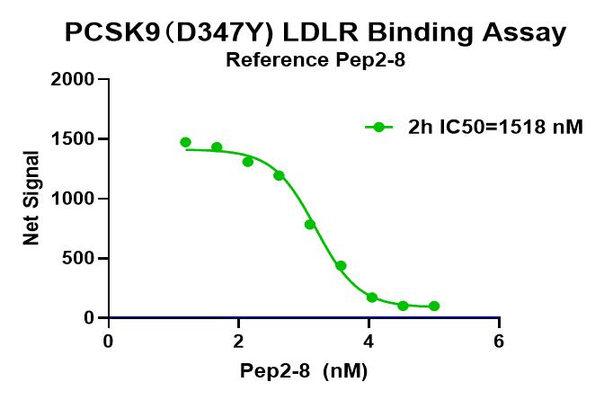
[Example Data]
The following data is illustrative and may vary based on instrumentation.
Note: Recommended microplate (384-well, white, shallow well)
```