UniOne® TR-FRET Human cGMP Detection Kit
UniOne® TR-FRET Human cGMP Detection Kit
Product Details
Product Details
Product Specification
| Host | Human |
| Stability & Storage | -80℃ |
Background
The kit employs homogeneous time-resolved fluorescence technology (TR-FRET), enabling precise measurement of cellular cGMP production. This method provides a high-throughput format for simple and rapid screening of molecules that modulate phosphodiesterase activity.
As illustrated, biotin-labeled cGMP can bind to streptavidin (SA), while the Eu-labeled anti-cGMP antibody (TR-FRET donor) and the Ac-labeled anti-Tag2 antibody (TR-FRET acceptor) bind to cGMP and biotin, respectively. The proximity of the donor and acceptor antibodies allows the excitation of the donor antibody to trigger fluorescence resonance energy transfer (FRET) to the acceptor antibody, resulting in a specific 665 nm emission signal. When cGMP is present in cell samples or standard curves, it competes with biotin-labeled cGMP for binding to the Eu-labeled anti-cGMP antibody, thereby reducing the FRET signal. This specific signal is proportional to the degree of interaction between the Eu-labeled anti-cGMP antibody, biotin-labeled cGMP, and SA. This homogeneous assay is simple to perform and requires no washing steps.

Components
Component |
Concentration |
100T |
500T |
2500T |
10000T |
Storage Temperature |
Biotin-cGMP protein |
100× |
5μL |
12.5μL |
62.5μL |
250μL |
-80℃ |
cGMP calibrator |
500μM |
5μL |
5μL |
25μL |
100μL |
-80℃ |
Anti-cGMP Eu antibody |
100× |
5μL |
25μL |
125μL |
500μL |
-80℃ |
SA-Ac |
12.5× |
20μL |
100μL |
500μL |
2mL |
-80℃ |
Lysis & Detect Buffer |
1× |
2mL |
10mL |
50mL |
200mL |
-80℃ |
Stimulation Buffer (without IBMX) |
1× |
2mL |
10mL |
50mL |
200mL |
-80℃ |
Protocol
Here is the English translation of the provided content while preserving the exact HTML structure and formatting: ```html
1. Reagent Preparation
1.1 Thaw all reagents to room temperature before use. The reaction system for the 384-well plate is 20μL (reagent volumes are shown in the table below). Calculate the required volume before preparation. The following preparation is for reference only, using 500 reactions as an example.
Table 1. Reagent Preparation and Volumes
Reagent Name |
Preparation |
Volume per Well (μL) |
Anti-cGMP Eu antibody |
Take 25μL of Anti-cGMP Eu antibody stock solution and dilute to 2.5mL with Lysis & Detect Buffer, mix well. |
5μL |
cGMP Ac Mix |
Take 12.5μL of Biotin-cGMP protein stock solution and dilute to 1.25mL with Lysis & Detect Buffer, mix well; take 100μL of SA-Ac stock solution and add to 1.25mL of Lysis & Detect Buffer, mix well. Mix the diluted Biotin-cGMP protein and SA-Ac in a 1:1 ratio to prepare the cGMP Ac Mix. |
5μL |
1.2 Gradient Dilution of Test Samples
Prepare according to experimental requirements. The following is for reference only.
Using cGMP calibrator as an example, the dilution buffer is Stimulation Buffer. Adjust the sample concentration as needed.
Table 2. cGMP Calibrator Preparation
|
Final Concentration (nM) |
Preparation Concentration (nM) |
Preparation Method |
| 1 | 1,250.000 |
5,000.000 |
1μL of 500μM stock + 99μL Stimulation Buffer |
| 2 | 416.667 |
1,666.667 |
20μL of ① + 40μL Stimulation Buffer |
| 3 | 138.889 |
555.556 |
20μL of ② + 40μL Stimulation Buffer |
| 4 | 46.296 |
185.185 |
20μL of ③ + 40μL Stimulation Buffer |
| 5 | 15.432 |
61.728 |
20μL of ④ + 40μL Stimulation Buffer |
| 6 | 5.144 |
20.576 |
20μL of ⑤ + 40μL Stimulation Buffer |
| 7 | 1.715 |
6.859 |
20μL of ⑥ + 40μL Stimulation Buffer |
| 8 | 0.572 |
2.286 |
20μL of ⑦ + 40μL Stimulation Buffer |
| 9 | 0.191 |
0.762 |
20μL of ⑧ + 40μL Stimulation Buffer |
| 10 | 0.064 |
0.254 |
20μL of ⑨ + 40μL Stimulation Buffer |
| 11 | 0.021 |
0.085 |
20μL of ⑩ + 40μL Stimulation Buffer |
Blank |
0 |
0 |
40μL of 1× Stimulation Buffer |
1.3 Pre-Treatment for Cell-Based Experiments
① Stimulation Buffer is used to dilute cells and stimulant drugs. The Stimulation Buffer provided in the kit does not contain IBMX. If the experiment aims to quantify the final concentration of cGMP in cells, it is necessary to add an appropriate concentration of IBMX to prevent cGMP degradation. If the experiment aims to evaluate the performance of PDE inhibitors, IBMX should not be added.
② Before conducting formal experiments, determine the number of cells per well and the incubation time with drugs based on cell and drug characteristics to ensure that the cGMP levels produced by cells fall within the linear range of the calibration curve.
2. Sample Addition and Controls
2.1 cGMP Standard Curve: 5μL of gradient-diluted cGMP reference, 5μL Stimulation Buffer, 5μL Anti-cGMP Eu antibody working solution, incubate for 10min, then add 5μL cGMP Ac Mix.
2.2 Test Samples: 5μL of gradient-diluted test sample, 5μL Stimulation Buffer, 5μL Anti-cGMP Eu antibody working solution, incubate for 10min, then add 5μL cGMP Ac Mix.
2.3 Cell-Based Experiments: Based on pre-experiment results, add 5μL of cells, 5μL of gradient-diluted drug, and incubate for the determined time. After incubation, add 5μL Anti-cGMP Eu antibody working solution, incubate at room temperature for 10min, then add 5μL cGMP Ac Mix.
2.4 Negative Control (NC): 5μL Stimulation Buffer instead of test sample or 5μL of untreated cells.
|
cGMP Standard Curve |
Test Samples |
Cell Experiments |
Negative Control (NC) |
|
Step 1
|
5μL cGMP calibrator |
5μL test sample |
5μL cells |
5μL Stimulation Buffer or 5μL cells |
5μL Stimulation Buffer |
5μL Stimulation Buffer |
5μL drug※ |
5μL Stimulation Buffer |
|
5μL Anti-cGMP Eu antibody working solution | ||||
Incubate at room temperature for 10min | ||||
Step 2 |
5μL cGMP Ac Mix |
|||
Seal the plate, centrifuge at 1000rpm for 1min to mix, incubate at room temperature for 120min, then read the plate. | ||||
•The incubation time of cells with drugs needs to be determined experimentally. Add subsequent reagents after drug stimulation of cells.
3.Detection
Measure using a TR-FRET-compatible microplate reader. Excitation wavelength: 320/340nm, emission wavelengths: 620nm and 665nm.
【Result Calculation】
1) Calculate the signal value (Ratio): Multiply the 665nm fluorescence signal by the 620nm fluorescence signal and then by 10,000.
Ratio = (665/620) × 10,000
2) Calculate CV (%):
CV (%) = Standard Deviation / Mean Ratio × 100%
【Data Example】
The following data is for reference only and may vary depending on the plate reader used.
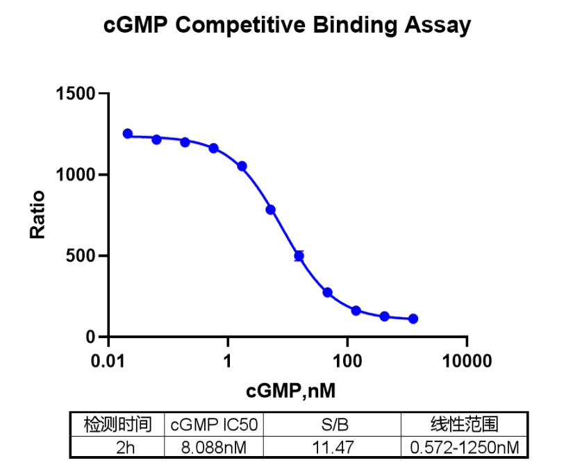
3 /3Note: *Data is for reference only. Recommended microplate: 384-well plate, white, shallow well.