WB result of Rabbit mAb IgG Isotype Control Primary antibody: Rabbit mAb IgG Isotype Control at 1/1000 dilution Lane 1: HeLa whole cell lysate 20 µg Lane 2: THP-1 whole cell lysate 20 µg Secondary antibody: Goat Anti-Rabbit IgG, (H+L), HRP conjugated at 1/10000 dilution
Rabbit mAb IgG Isotype Control (S-240-1)
Rabbit mAb IgG Isotype Control (S-240-1)
Price:
Regular price
$45 USD
Regular price
Sale price
$45 USD
Unit price
per
For shipping services or bulk orders, you may request a quotation.
Secure checkout with

View full details
Product Details
Product Details
Product Specification
| Host | Rabbit |
| Immunogen | Recombinant Protein |
| Clone Number | S-240-1 |
| Antibody Type | Rabbit mAb |
| Application | WB, IHC-P, FCM, IP |
| Purification | Protein A |
| Concentration | 0.5 mg/ml |
| Conjugation | Unconjugated |
| Physical Appearance | Liquid |
| Storage Buffer | PBS, 40% Glycerol, 0.05% BSA, 0.03% Proclin 300 |
| Stability & Storage | 12 months from date of receipt / reconstitution, -20 °C as supplied |
Background
Isotype control antibodies, to estimate the nonspecific binding of target. Use at concentrations comparable to those of the specific antibody of interest.
Picture
Picture
Western Blot
WB result of Rabbit mAb IgG Isotype Control Primary antibody: Rabbit mAb IgG Isotype Control at 1/1000 dilution Lane 1: NIH/3T3 whole cell lysate 20 µg Lane 2: mouse testis lysate 20 µg Secondary antibody: Goat Anti-Rabbit IgG, (H+L), HRP conjugated at 1/10000 dilution
WB result of Rabbit mAb IgG Isotype Control Primary antibody: Rabbit mAb IgG Isotype Control at 1/1000 dilution Lane 1: C6 whole cell lysate 20 µg Lane 2: rat testis lysate 20 µg Secondary antibody: Goat Anti-Rabbit IgG, (H+L), HRP conjugated at 1/10000 dilution
FC
Flow cytometric analysis of mouse primary splenocytes labeling Rabbit IgG isotype control at 1/50 dilution (1 μg) / (left panel) compared with CD8α antibody at 1/50 (1 μg) dilution (S0B0034) / (right panel). Goat Anti-Rabbit IgG Alexa Fluor 488 was used as the secondary antibody. Then cells were stained with CD4 - Alexa Fluor 647 separately. CD4 and CD8α are mutually exclusive expressed in mouse primary splenocytes. Gated on total viable cells.
Flow cytometric analysis of 4% PFA fixed 90% methanol permeabilized HeLa (Human cervix adenocarcinoma epithelial cell) labeling Rabbit IgG isotype control at 1/50 dilution (1 μg) / (left panel) compared with Stat6 antibody at 1/50 (1 μg) dilution (S0B0085) / (right panel). Goat Anti-Rabbit IgG Alexa Fluor 488 was used as the secondary antibody.
IP
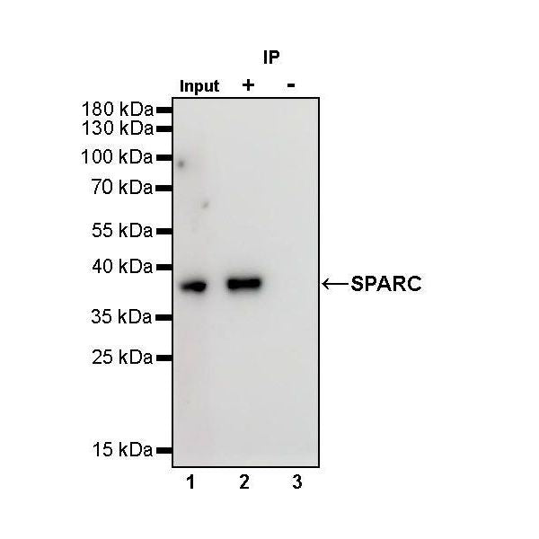
SPARC Rabbit mAb at 1/50 dilution (1 µg) immunoprecipitating SPARC in 0.4 mg A375 whole cell lysate.
Western blot was performed on the immunoprecipitate using SPARC Rabbit mAb at 1/1000 dilution.
Secondary antibody (HRP) for IP was used at 1/400 dilution.
Lane 1: A375 whole cell lysate 20 µg (Input)
Lane 2: SPARC Rabbit mAb IP in A375 whole cell lysate
Lane 3: Rabbit mAb IgG Isotype Control IP in A375 whole cell lysate
Predicted MW: 35 kDa
Observed MW: 37 kDa
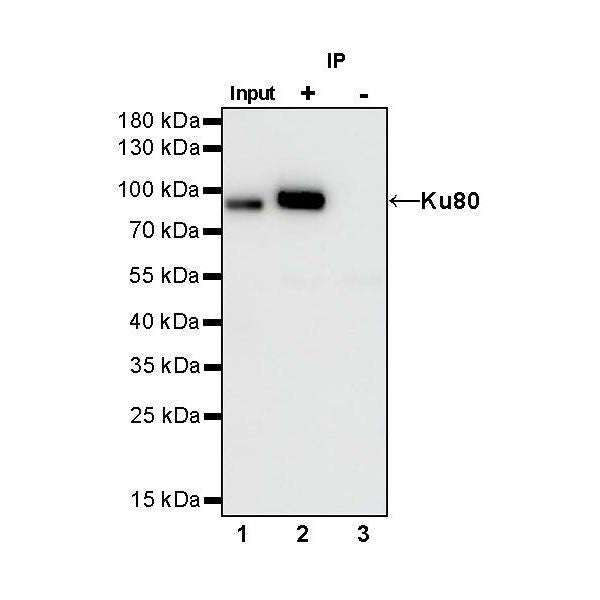
Ku80 Rabbit mAb at 1/50 dilution (1 µg) immunoprecipitating Ku80 in 0.4 mg HeLa whole cell lysate.
Western blot was performed on the immunoprecipitate using Ku80 Rabbit mAb at 1/1000 dilution.
Secondary antibody (HRP) for IP was used at 1/400 dilution.
Lane 1: HeLa whole cell lysate 20 µg (Input)
Lane 2: Ku80 Rabbit mAb IP in HeLa whole cell lysate
Lane 3: Rabbit mAb IgG Isotype Control IP in HeLa whole cell lysate
Predicted MW: 83 kDa
Observed MW: 86 kDa
Immunohistochemistry
IHC shows negative staining in paraffin-embedded human cerebral cortex. Rabbit mAb IgG Isotype Control was used at 1/500 dilution, followed by a HRP Polymer for Mouse & Rabbit IgG (ready to use). Counterstained with hematoxylin. Heat mediated antigen retrieval with Tris/EDTA buffer pH9.0 was performed before commencing with IHC staining protocol.
IHC shows negative staining in paraffin-embedded human kidney. Rabbit mAb IgG Isotype Control was used at 1/500 dilution, followed by a HRP Polymer for Mouse & Rabbit IgG (ready to use). Counterstained with hematoxylin. Heat mediated antigen retrieval with Tris/EDTA buffer pH9.0 was performed before commencing with IHC staining protocol.
IHC shows negative staining in paraffin-embedded human tonsil. Rabbit mAb IgG Isotype Control was used at 1/500 dilution, followed by a HRP Polymer for Mouse & Rabbit IgG (ready to use). Counterstained with hematoxylin. Heat mediated antigen retrieval with Tris/EDTA buffer pH9.0 was performed before commencing with IHC staining protocol.
IHC shows negative staining in paraffin-embedded human breast cancer. Rabbit mAb IgG Isotype Control was used at 1/500 dilution, followed by a HRP Polymer for Mouse & Rabbit IgG (ready to use). Counterstained with hematoxylin. Heat mediated antigen retrieval with Tris/EDTA buffer pH9.0 was performed before commencing with IHC staining protocol.
IHC shows negative staining in paraffin-embedded human colon cancer. Rabbit mAb IgG Isotype Control was used at 1/500 dilution, followed by a HRP Polymer for Mouse & Rabbit IgG (ready to use). Counterstained with hematoxylin. Heat mediated antigen retrieval with Tris/EDTA buffer pH9.0 was performed before commencing with IHC staining protocol.
IHC shows negative staining in paraffin-embedded mouse cerebral cortex. Rabbit mAb IgG Isotype Control was used at 1/500 dilution, followed by a HRP Polymer for Mouse & Rabbit IgG (ready to use). Counterstained with hematoxylin. Heat mediated antigen retrieval with Tris/EDTA buffer pH9.0 was performed before commencing with IHC staining protocol.
IHC shows negative staining in paraffin-embedded rat spleen. Rabbit mAb IgG Isotype Control was used at 1/500 dilution, followed by a HRP Polymer for Mouse & Rabbit IgG (ready to use). Counterstained with hematoxylin. Heat mediated antigen retrieval with Tris/EDTA buffer pH9.0 was performed before commencing with IHC staining protocol.