WB result of Neurofilament M Rabbit mAb
Primary antibody: Nucleophosmin/NPM Rabbit mAb at 1/1000 dilution
Lane 1: Jurkat whole cell lysate 20 µg
Lane 2: HeLa whole cell lysate 20 µg
Secondary antibody: Goat Anti-Rabbit IgG, (H+L), HRP conjugated at 1/10000 dilution
Predicted MW: 33 kDa
Observed MW: 38 kDa
Nucleophosmin/NPM Recombinant Rabbit mAb (S-R231)
Nucleophosmin/NPM Recombinant Rabbit mAb (S-R231)
Price:
Regular price
$100 USD
Regular price
Sale price
$100 USD
Unit price
per
For shipping services or bulk orders, you may request a quotation.
Secure checkout with

View full details
Product Details
Product Details
Product Specification
| Host | Rabbit |
| Antigen | Nucleophosmin/NPM |
| Synonyms | Nucleolar phosphoprotein B23, Nucleolar protein NO38, Numatrin, NPM1 |
| Location | Nucleus, Nucleolus |
| Accession | P06748 |
| Clone Number | S-R231 |
| Antibody Type | Recombinant mAb |
| Application | WB, IHC-P, ICC, ICFCM, IP |
| Reactivity | Hu, Ms, Rt |
| Purification | Protein A |
| Concentration | 0.5 mg/ml |
| Conjugation | Unconjugated |
| Physical Appearance | Liquid |
| Storage Buffer | PBS, 40% Glycerol, 0.05%BSA, 0.03% Proclin 300 |
| Stability & Storage | 12 months from date of receipt / reconstitution, -20 °C as supplied |
Dilution
| application | dilution | species |
| WB | 1:1000 | null |
| IHC | 1:250 | null |
| ICFCM | 1:500 | null |
| IP | 1:50 | null |
| ICC | 1:500 | null |
Background
Nucleophosmin (NPM), also known as nucleolar phosphoprotein B23 or numatrin, is a protein that in humans is encoded by the NPM1 gene. NPM1 is associated with nucleolar ribonucleoprotein structures and binds single-stranded and double-stranded nucleic acids, but it binds preferentially G-quadruplex forming nucleic acids. It is involved in the biogenesis of ribosomes and may assist small basic proteins in their transport to the nucleolus. Its regulation through SUMOylation (by SENP3 and SENP5) is another facet of the protein's regulation and cellular functions. It is located in the nucleolus, but it can be translocated to the nucleoplasm in case of serum starvation or treatment with anticancer drugs. The protein is phosphorylated.
Picture
Picture
Western Blot
WB result of Nucleophosmin/NPM Rabbit mAb
Primary antibody: Nucleophosmin/NPM Rabbit mAb at 1/1000 dilution
Lane 1: NIH/3T3 whole cell lysate 20 µg
Secondary antibody: Goat Anti-Rabbit IgG, (H+L), HRP conjugated at 1/10000 dilution
Predicted MW: 33 kDa
Observed MW: 38 kDa
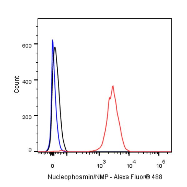
FC
Flow cytometric analysis of 4% PFA fixed 90% methanol permeabilized Jurkat (Human T cell leukemia T lymphocyte) cells labeling Nucleophosmin/NMP at 1/500 dilution (0.1 μg) / (red) compared with a rabbit monoclonal IgG isotype control (black) and an unlabeled control (cells without incubation with primary antibody and secondary antibody) (Blue). Goat Anti - Rabbit IgG Alexa Fluor® 488 was used as the secondary antibody.
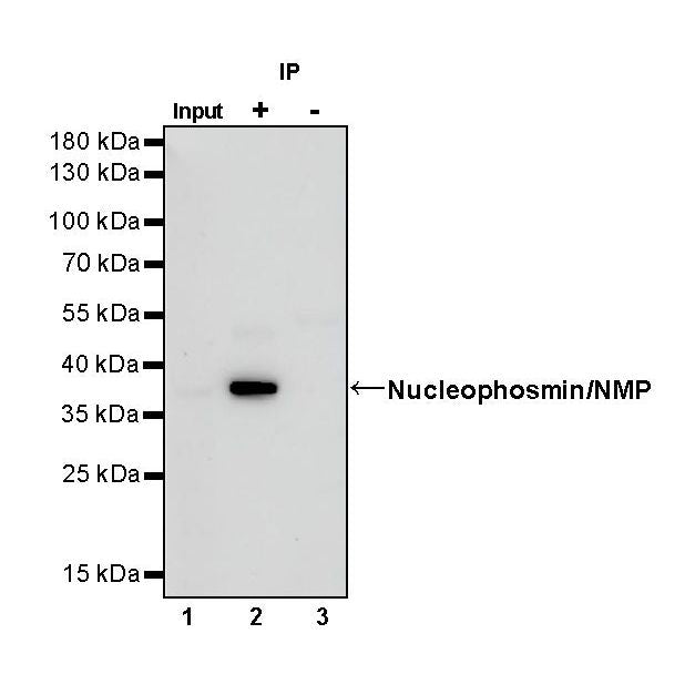
IP
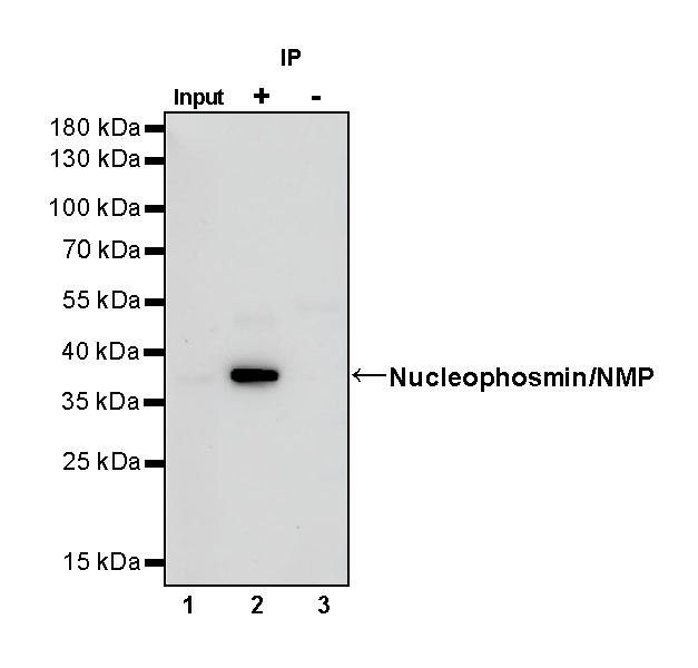
Nucleophosmin/NMP Rabbit mAb at 1/50 dilution (1 µg) immunoprecipitating Nucleophosmin/NMP in 0.4 mg Jurkat whole cell lysate.
Western blot was performed on the immunoprecipitate using Nucleophosmin/NMP Rabbit mAb at 1/1000 dilution.
Secondary antibody (HRP) for IP was used at 1/400 dilution.
Lane 1: Jurkat whole cell lysate 20 µg (Input)
Lane 2: Nucleophosmin/NMP Rabbit mAb IP in Jurkat whole cell lysate
Lane 3: Rabbit monoclonal IgG IP in Jurkat whole cell lysate
Predicted MW: 33 kDa
Observed MW: 38 kDa
Immunohistochemistry
IHC shows positive staining in paraffin-embedded human stomach. Anti-Nucleophosmin/NPM antibody was used at 1/250 dilution, followed by a HRP Polymer for Mouse & Rabbit IgG (ready to use). Counterstained with hematoxylin. Heat mediated antigen retrieval with Tris/EDTA buffer pH9.0 was performed before commencing with IHC staining protocol.
IHC shows positive staining in paraffin-embedded human testis. Anti-Nucleophosmin/NPM antibody was used at 1/250 dilution, followed by a HRP Polymer for Mouse & Rabbit IgG (ready to use). Counterstained with hematoxylin. Heat mediated antigen retrieval with Tris/EDTA buffer pH9.0 was performed before commencing with IHC staining protocol.
IHC shows positive staining in paraffin-embedded human ovarian cancer. Anti-Nucleophosmin/NMP antibody was used at 1/250 dilution, followed by a HRP Polymer for Mouse & Rabbit IgG (ready to use). Counterstained with hematoxylin. Heat mediated antigen retrieval with Tris/EDTA buffer pH9.0 was performed before commencing with IHC staining protocol.
IHC shows positive staining in paraffin-embedded human gastric cancer. Anti-Nucleophosmin/NMP antibody was used at 1/250 dilution, followed by a HRP Polymer for Mouse & Rabbit IgG (ready to use). Counterstained with hematoxylin. Heat mediated antigen retrieval with Tris/EDTA buffer pH9.0 was performed before commencing with IHC staining protocol.
IHC shows positive staining in paraffin-embedded human pancreas cancer. Anti-Nucleophosmin/NPM antibody was used at 1/250 dilution, followed by a HRP Polymer for Mouse & Rabbit IgG (ready to use). Counterstained with hematoxylin. Heat mediated antigen retrieval with Tris/EDTA buffer pH9.0 was performed before commencing with IHC staining protocol.
IHC shows positive staining in paraffin-embedded mouse colon. Anti-Nucleophosmin/NPM antibody was used at 1/250 dilution, followed by a HRP Polymer for Mouse & Rabbit IgG (ready to use). Counterstained with hematoxylin. Heat mediated antigen retrieval with Tris/EDTA buffer pH9.0 was performed before commencing with IHC staining protocol.
IHC shows positive staining in paraffin-embedded mouse liver. Anti-Nucleophosmin/NPM antibody was used at 1/250 dilution, followed by a HRP Polymer for Mouse & Rabbit IgG (ready to use). Counterstained with hematoxylin. Heat mediated antigen retrieval with Tris/EDTA buffer pH9.0 was performed before commencing with IHC staining protocol.
IHC shows positive staining in paraffin-embedded rat kidney. Anti-Nucleophosmin/NPM antibody was used at 1/250 dilution, followed by a HRP Polymer for Mouse & Rabbit IgG (ready to use). Counterstained with hematoxylin. Heat mediated antigen retrieval with Tris/EDTA buffer pH9.0 was performed before commencing with IHC staining protocol.
Immunocytochemistry
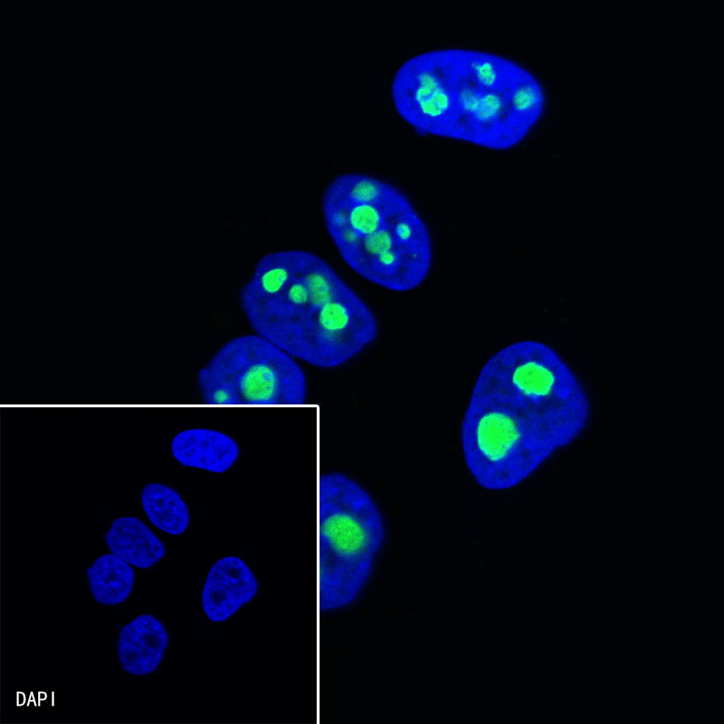
ICC shows positive staining in HeLa cells. Anti-Nucleophosmin/NMP antibody was used at 1/500 dilution (Green) and incubated overnight at 4°C. Goat polyclonal Antibody to Rabbit IgG - H&L (Alexa Fluor® 488) was used as secondary antibody at 1/1000 dilution. The cells were fixed with 4%PFA and permeabilized with 0.1% PBS-Triton X-100. Nuclei were counterstained with DAPI (Blue).