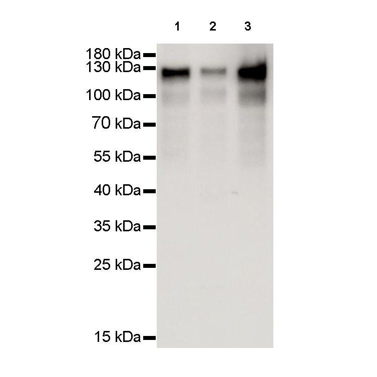
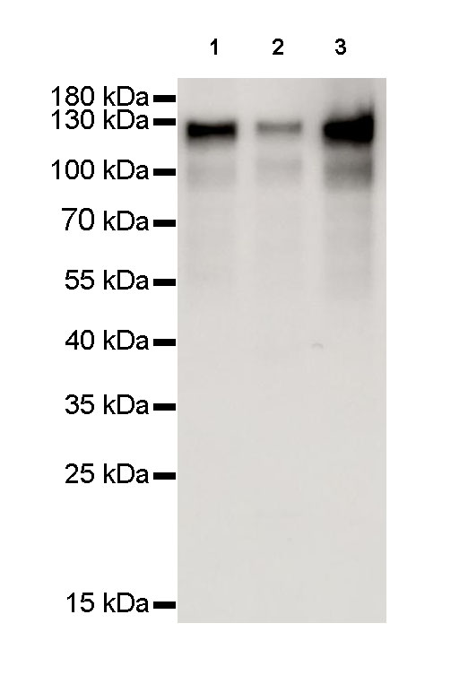
WB result of MCM2 Rabbit mAb
Primary antibody : MCM2 Rabbit mAb at 1/8000 dilution
Lane 1 : Hela whole cell lysate 20 µg
Lane 2 : Ramos whole cell lysate 20 µg
Lane 3 : Molt-4 whole cell lysate 20 µg
Secondary antibody: Goat Anti-Rabbit IgG, (H+L), HRP conjugated at 1/10000 dilution
Predicted MW: 125 kDa
Observed MW: 125 kDa
Exposure time: 0.5 seconds
MCM2 Recombinant Rabbit mAb (SDT-018-68-2)
MCM2 Recombinant Rabbit mAb (SDT-018-68-2)
Price:
Regular price
$100 USD
Regular price
Sale price
$100 USD
Unit price
per
For shipping services or bulk orders, you may request a quotation.
Secure checkout with

View full details
Product Details
Product Details
Product Specification
| Host | Rabbit |
| Antigen | MCM2 |
| Synonyms | BM28, CCNL1, CDCL1, Minichromosome maintenance protein 2 homolog, Nuclear protein BM28 |
| Immunogen | Synthetic Peptide |
| Location | Nucleus |
| Accession | P49736 |
| Clone Number | SDT-018-68-2 |
| Antibody Type | Rabbit mAb |
| Isotype | IgG |
| Application | WB, IHC-P, ICC, ICFCM |
| Reactivity | Hu, Ms |
| Purification | Protein A |
| Research Area | Epigenetics |
| Concentration | 0.5mg/ml |
| Molecular Weight | 125kDa |
| Conjugation | Unconjugated |
| Physical Appearance | Liquid |
| Storage Buffer | PBS, 40% Glycerol, 0.05%BSA, 0.03% Proclin 300 |
| Stability & Storage | 12 months from date of receipt / reconstitution, -20 °C as supplied |
Dilution
| application | dilution | species |
| ICFCM | 1:500 | |
| WB | 1:8000 | |
| IHC-P | 1:1000 | |
| ICC | 1:500 |
Background
MCM2 is one of the highly conserved mini-chromosome maintenance proteins (MCM) that are involved in the initiation of eukaryotic genome replication. The hexameric protein complex formed by MCM proteins is a key component of the pre-replication complex (pre-RC) and may be involved in the formation of replication forks and in the recruitment of other DNA replication related proteins. MCM2 forms a complex with MCM4, 6, and 7, and has been shown to regulate the helicase activity of the complex. MCM2 is phosphorylated, and thus regulated by protein kinases CDC2 and CDC7.
Picture
Picture
Western Blot
FC
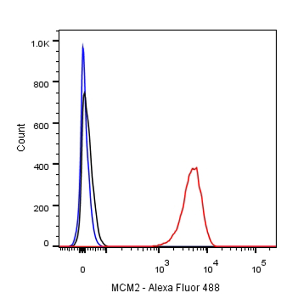
Flow cytometric analysis of Jurkat cells labelling MCM2 antibody at 1/500 (0.1 μg) dilution/ (red) compared with a Rabbit monoclonal IgG (Black) isotype control and an unlabelled control (cells without incubation with primary antibody and secondary antibody) (Blue). Goat Anti-Rabbit IgG Alexa Fluor® 488 was used as the secondary antibody.
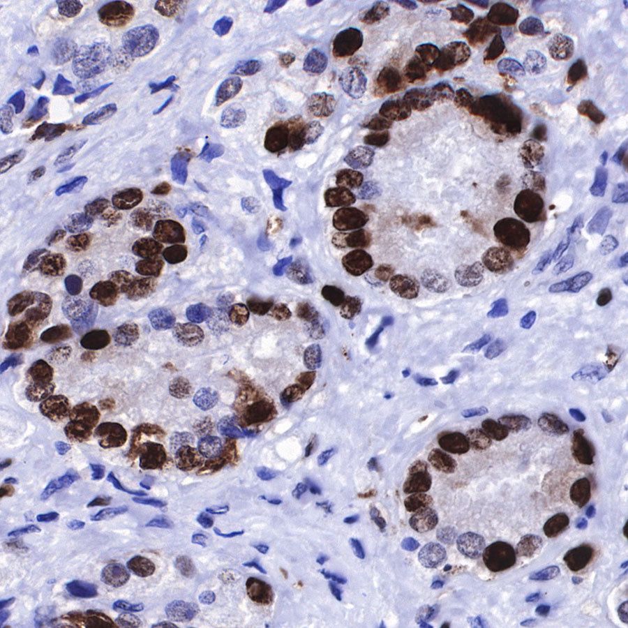
Immunohistochemistry

IHC show nucleus staining in paraffin-embedded human tonsil. Anti-MCM2 antibody was used at 1/1000 dilution, followed by a Goat Anti-Rabbit IgG H&L (HRP) ready to use. Counterstained with hematoxylin.
Heat mediated antigen retrieval with Tris/EDTA buffer pH9.0 was performed before commencing with IHC staining protocol.
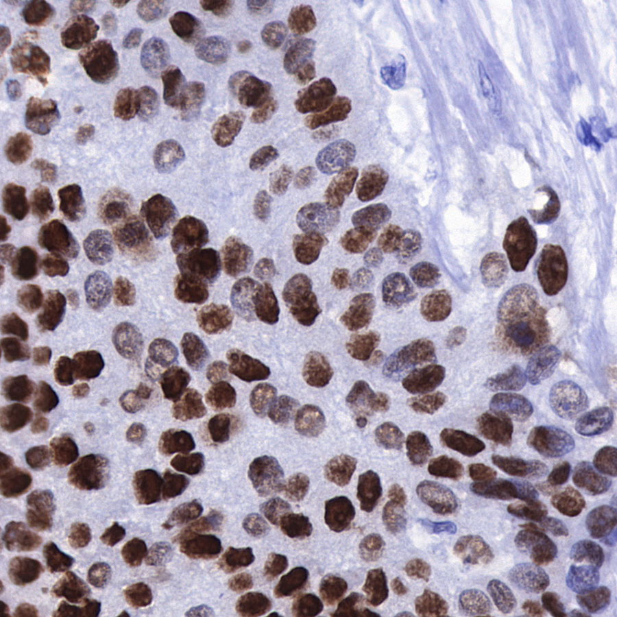
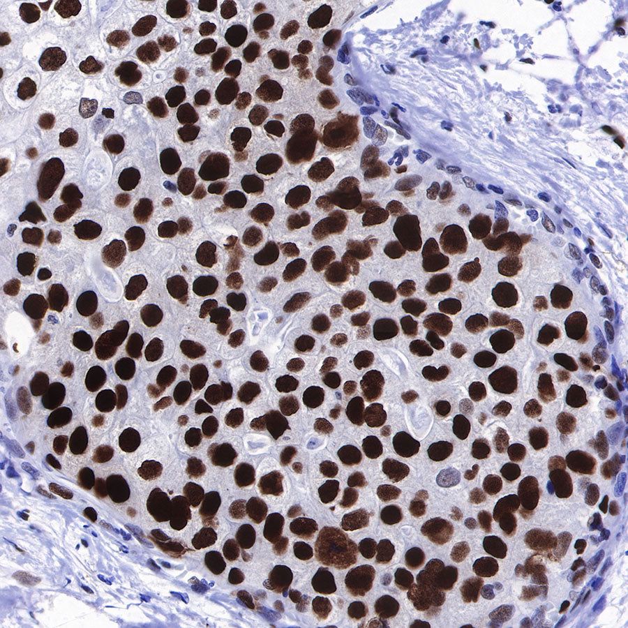
IHC show nucleus staining in paraffin-embedded human bladder cancer. Anti-MCM2 antibody was used at 1/1000 dilution, followed by a Goat Anti-Rabbit IgG H&L (HRP) ready to use. Counterstained with hematoxylin.
Heat mediated antigen retrieval with Tris/EDTA buffer pH9.0 was performed before commencing with IHC staining protocol.

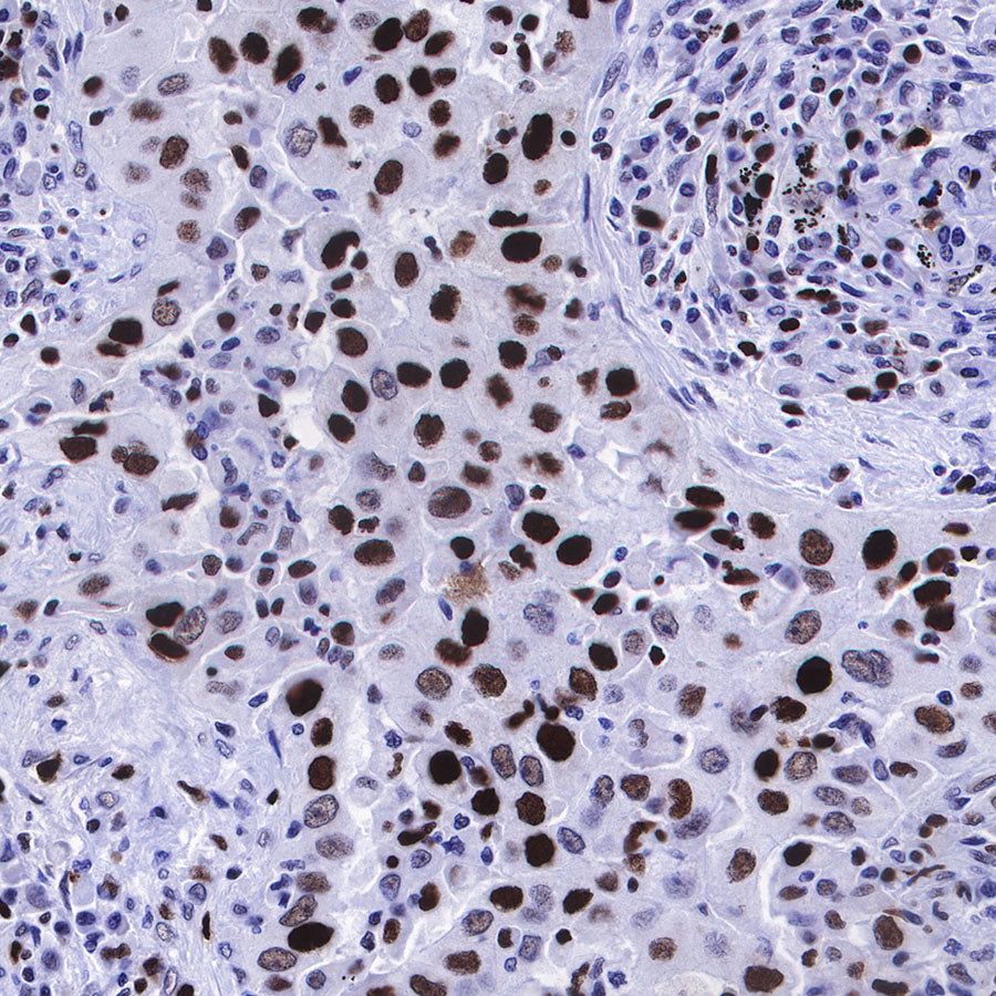
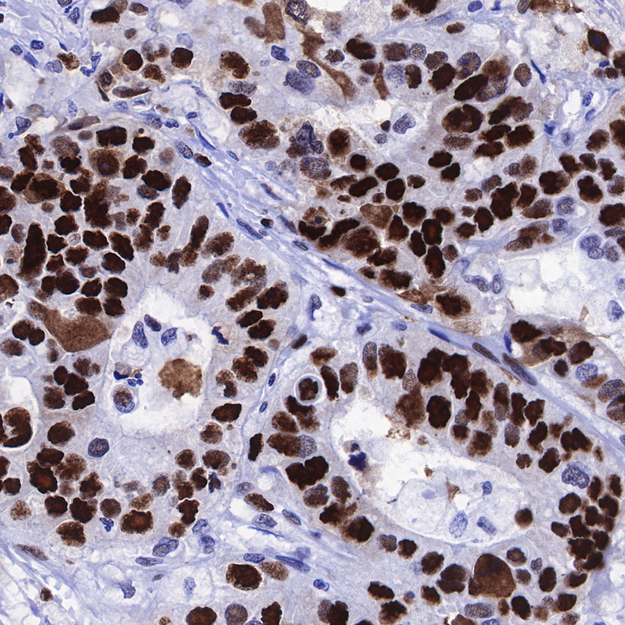
IHC show nucleus staining in paraffin-embedded human breast cancer. Anti-MCM2 antibody was used at 1/1000 dilution, followed by a Goat Anti-Rabbit IgG H&L (HRP) ready to use. Counterstained with hematoxylin.
Heat mediated antigen retrieval with Tris/EDTA buffer pH9.0 was performed before commencing with IHC staining protocol.
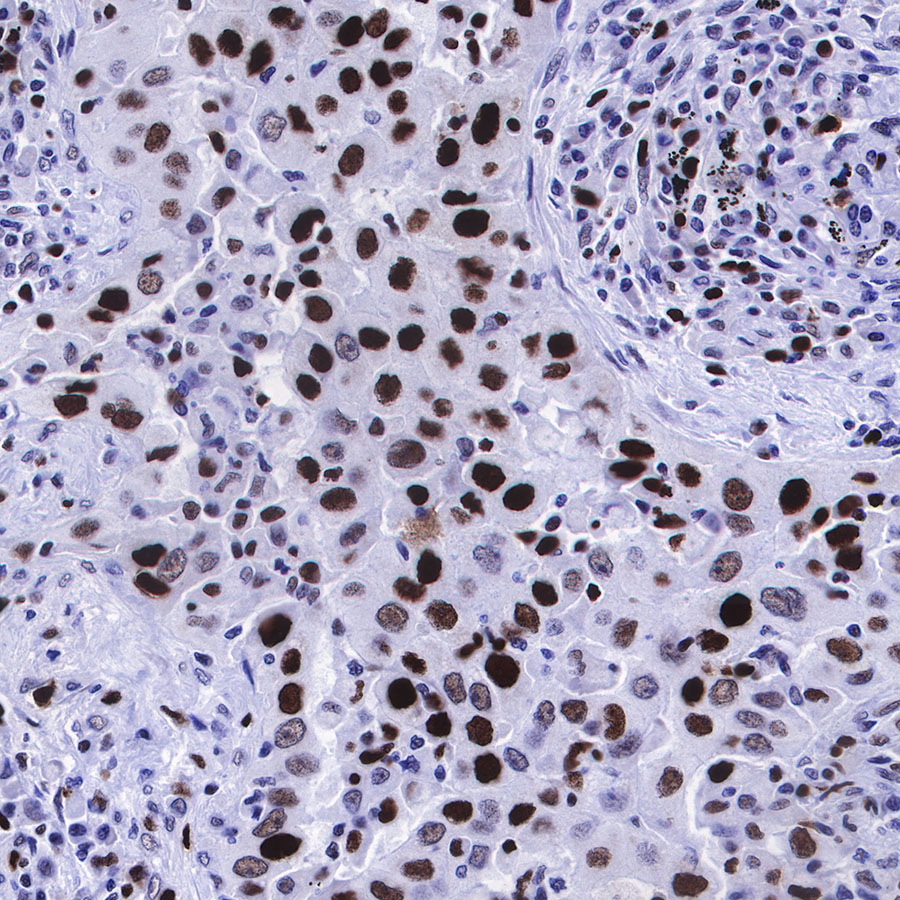
IHC show nucleus staining in paraffin-embedded human gastric cancer. Anti-MCM2 antibody was used at 1/1000 dilution, followed by a Goat Anti-Rabbit IgG H&L (HRP) ready to use. Counterstained with hematoxylin.
Heat mediated antigen retrieval with Tris/EDTA buffer pH9.0 was performed before commencing with IHC staining protocol.
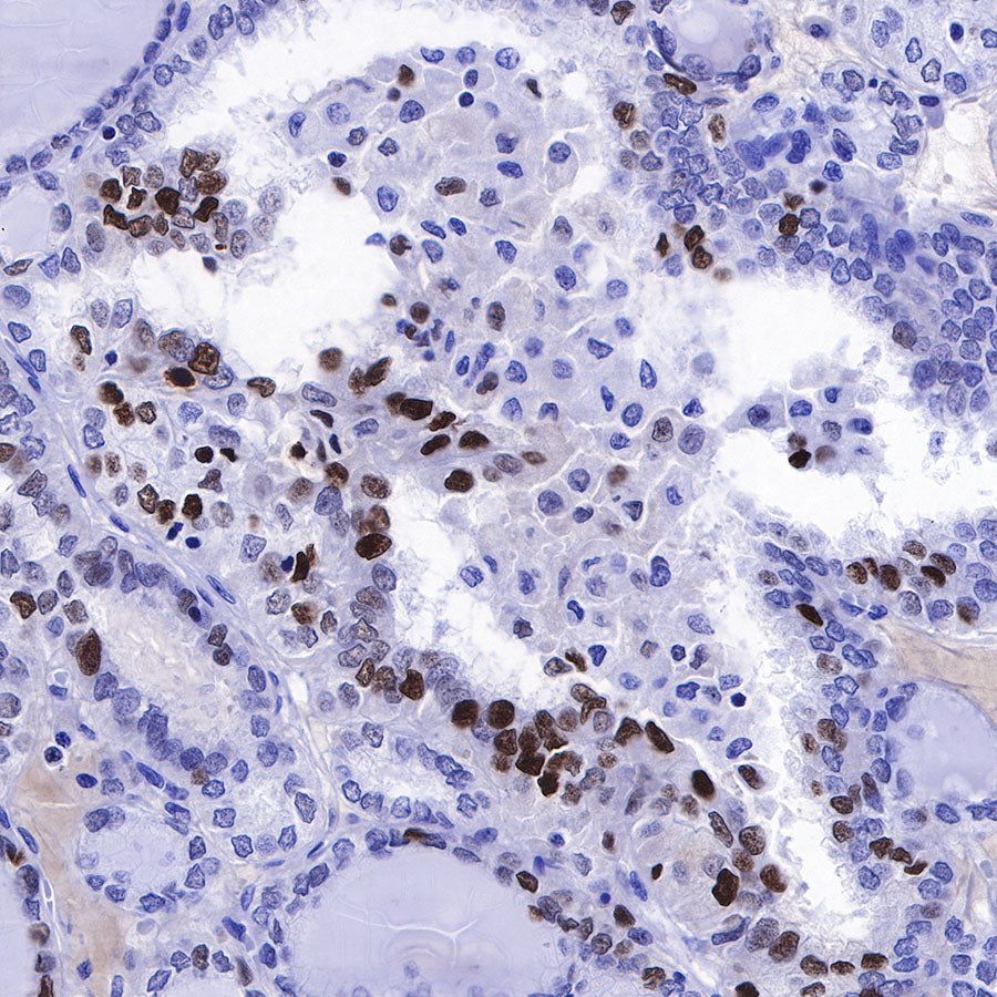
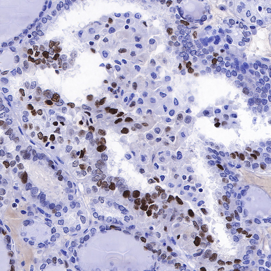
IHC show nucleus staining in paraffin-embedded human lung adenocarcinoma. Anti-MCM2 antibody was used at 1/1000 dilution, followed by a Goat Anti-Rabbit IgG H&L (HRP) ready to use. Counterstained with hematoxylin.
Heat mediated antigen retrieval with Tris/EDTA buffer pH9.0 was performed before commencing with IHC staining protocol.

IHC show nucleus staining in paraffin-embedded human pancreatic cancer. Anti-MCM2 antibody was used at 1/1000 dilution, followed by a Goat Anti-Rabbit IgG H&L (HRP) ready to use. Counterstained with hematoxylin.
Heat mediated antigen retrieval with Tris/EDTA buffer pH9.0 was performed before commencing with IHC staining protocol.
IHC show nucleus staining in paraffin-embedded human thyroid cancer. Anti-MCM2 antibody was used at 1/1000 dilution, followed by a Goat Anti-Rabbit IgG H&L (HRP) ready to use. Counterstained with hematoxylin.
Heat mediated antigen retrieval with Tris/EDTA buffer pH9.0 was performed before commencing with IHC staining protocol.
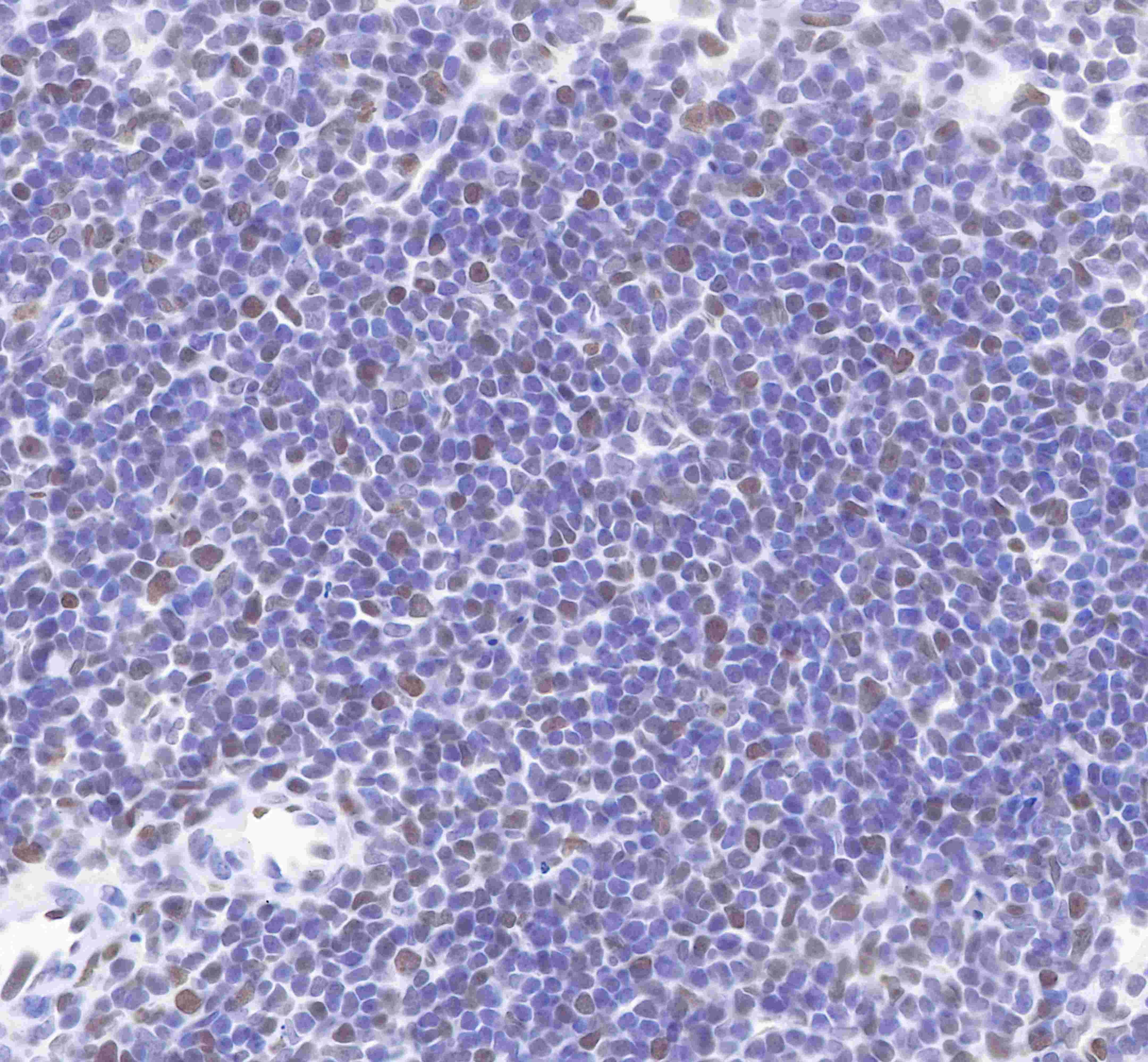
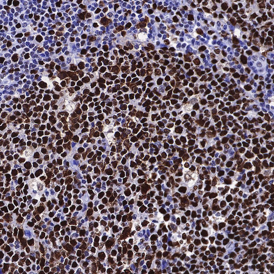
IHC show nucleus staining in paraffin-embedded mouse spleen. Anti-MCM2 antibody was used at 1/1000 dilution, followed by a Goat Anti-Rabbit IgG H&L (HRP) ready to use. Counterstained with hematoxylin.
Heat mediated antigen retrieval with Tris/EDTA buffer pH9.0 was performed before commencing with IHC staining protocol.
Immunocytochemistry
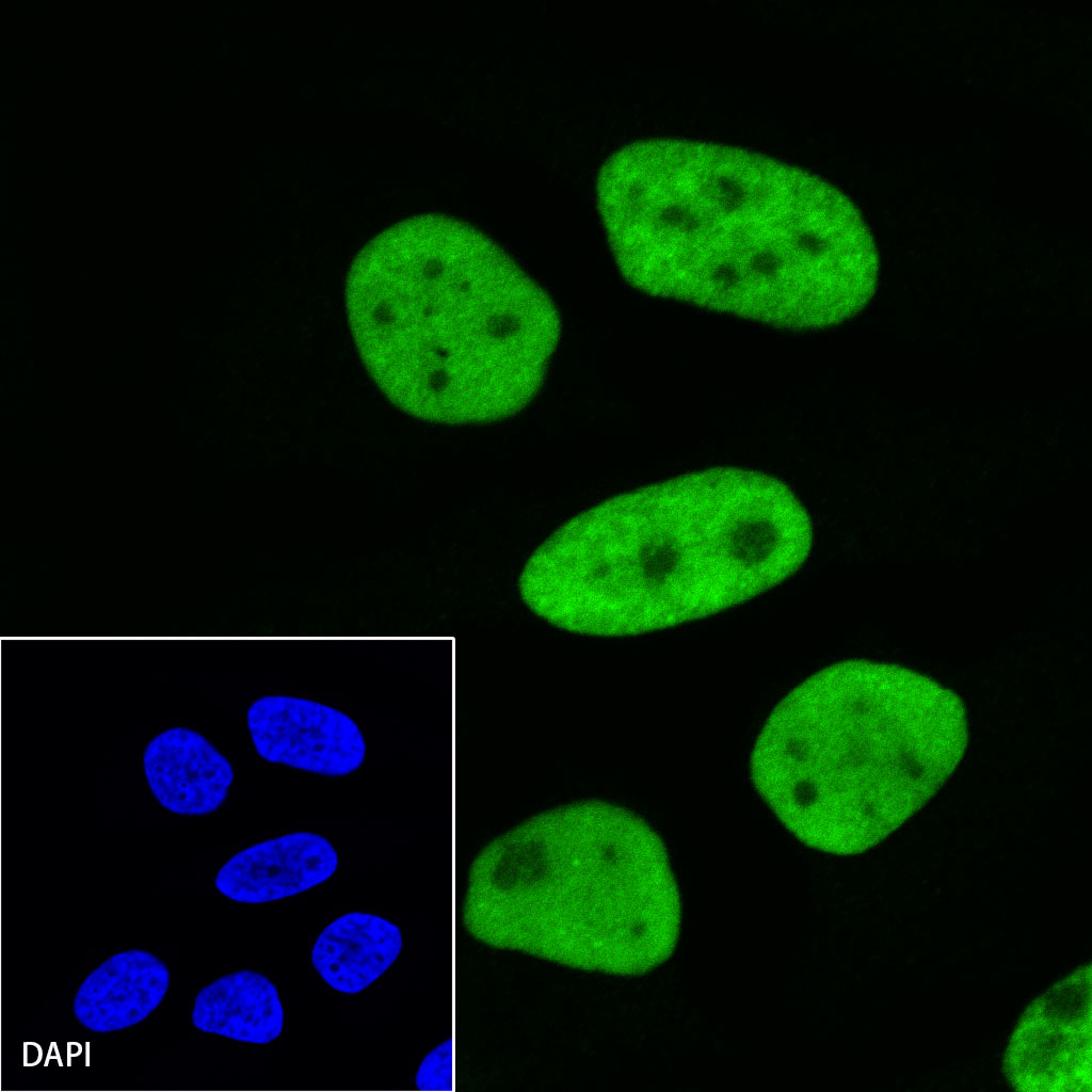
ICC shows positive staining in HeLa cells. Anti-MCM2 antibody was used at 1/500 dilution (Green) and incubated overnight at 4°C. Goat polyclonal Antibody to Rabbit IgG - H&L (Alexa Fluor® 488) was used as secondary antibody at 1/1000 dilution. The cells were fixed with 100% ice-cold methanol and permeabilized with 0.1% PBS-Triton X-100. Nuclei were counterstained with DAPI (Blue).