| Flow cytometric analysis of CD27 expression on human peripheral blood. Human peripheral blood was stained with Brilliant Violet 421™ Mouse Anti-Human CD19 antibody and either Alexa Fluor® 647 Armenian hamster IgG Isotype Control (Left panel) or SDT Alexa Fluor® 647 Armenian hamster Anti-Mouse/Rat/Human CD27 Antibody (Right panel) at 5 μl/test. Flow cytometry and data analysis were performed using BD FACSymphony™ A1 and FlowJo™ software. |
Alexa Fluor® 647 Armenian hamster Anti-Mouse/Rat/Human CD27 Antibody (S-R620)
Alexa Fluor® 647 Armenian hamster Anti-Mouse/Rat/Human CD27 Antibody (S-R620)
Price:
Regular price
$230 USD
Regular price
Sale price
$230 USD
Unit price
per
For shipping services or bulk orders, you may request a quotation.
Secure checkout with

View full details
Product Details
Product Details
Product Specification
| Host | Armenian hamster |
| Antigen | CD27 |
| Synonyms | CD27 antigen; CD27L receptor; T-cell activation antigen CD27; T14; Tumor necrosis factor receptor superfamily member 7; TNFRSF7 |
| Accession | P26842、 P41272、 Q501W2 |
| Clone Number | S-R620 |
| Antibody Type | Recombinant mAb |
| Application | FCM |
| Reactivity | Hu, Ms, Rt |
| Positive Sample | Human PBMC, Mouse splenocytes |
| Purification | Protein G |
| Concentration | 0.2 mg/ml |
| Conjugation | Alexa Fluor® 647 |
| Physical Appearance | Liquid |
| Storage Buffer | PBS, 25% Glycerol, 1% BSA, 0.3% Proclin 300 |
| Stability & Storage | 12 months from date of receipt / reconstitution, 2 to 8 °C as supplied. |
Dilution
| application | dilution | species |
| FCM | 5 μl per million cells in 100μl volume | Hu, Ms |
Background
CD27 is a transmembrane protein that belongs to the tumor necrosis factor receptor superfamily (TNFRSF) and is primarily expressed on T cells, B cells, and natural killer (NK) cells. It activates NF-κB and JNK signaling pathways by binding to its ligand, CD70, thereby promoting the activation, proliferation, and differentiation of T cells. CD27 plays a crucial role in immune regulation, acting as an important co-stimulatory receptor for T and B cells, and is involved in modulating immune responses and the formation of memory cells. Additionally, CD27 is being studied as a potential target for anti-tumor immunotherapy.
Picture
Picture
FC
| Flow cytometric analysis of CD27 expression on C57BL/6 mouse splenocytes. C57BL/6 mouse splenocytes were stained with PE/Cy7 Rat Anti-Mouse CD19 antibody and either Alexa Fluor® 647 Armenian hamster IgG Isotype Control (Left panel) or SDT Alexa Fluor® 647 Armenian hamster Anti-Mouse/Rat/Human CD27 Antibody (Right panel) at 5 μl/test. Flow cytometry and data analysis were performed using BD FACSymphony™ A1 and FlowJo™ software. |
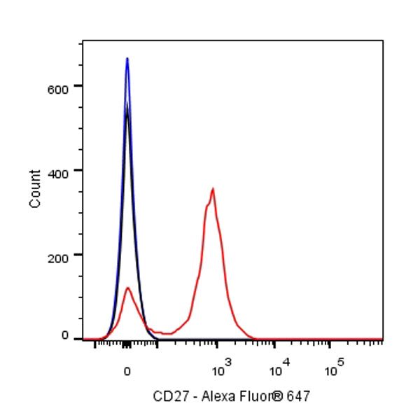
Flow cytometric analysis of CD27 expression on SD Rat splenocytes. SD Rat splenocytes were stained with either Alexa Fluor® 647 Armenian hamster IgG Isotype Control (Black line histogram) or SDT Alexa Fluor® 647 Armenian hamster Anti-Mouse/Rat/Human CD27 Antibody (Red line histogram) at 5 μl/test, cells without incubation with primary antibody and secondary antibody (Blue line histogram) was used as unlabelled control. Flow cytometry and data analysis were performed using BD FACSymphony™ A1 and FlowJo™ software.