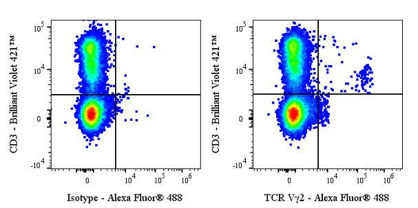
Flow cytometric analysis of C57BL/6 mouse splenocytes labeled with anti-mouse TCR Vγ2 antibody at 1/5000 dilution (0.1 μg) (right panel) compared to an Armenian hamster IgG Isotype Control (left panel). Goat Anti- Armenian hamster IgG Alexa Fluor® 488 was used as the secondary antibody. Then cells were stained with CD3 - Brilliant Violet 421™ antibody separately. Flow cytometry and data analysis were performed using Agilent NovoCyte Quanteon and FlowJo™ software.
Invivo anti-Mouse TCR Vγ2 Recombinant mAb
Invivo anti-Mouse TCR Vγ2 Recombinant mAb
Price:
Regular price
$120 USD
Regular price
Sale price
$120 USD
Unit price
per
For shipping services or bulk orders, you may request a quotation.
Secure checkout with

View full details
Product Details
Product Details
Product Specification
| Host | Armenian hamster |
| Antigen | TCR Vγ2 |
| Location | Cell membrane |
| Accession | A2J008 |
| Clone Number | UC3-10A6 |
| Antibody Type | Recombinant mAb |
| Isotype | Armenian hamster IgG |
| Isotype Control | Invivo Armenian Hamster IgG Isotype Control |
| Application | FCM, in vivo cell specific depletion |
| Reactivity | Ms |
| Positive Sample | C57BL/6 mouse splenocytes |
| Purification | Protein G |
| Concentration | 5 mg/ml |
| Purity | >95%(Determined by SDS-PAGE) |
| Endotoxin | <1EU/mg |
| Conjugation | Unconjugated |
| Physical Appearance | Liquid |
| Storage Buffer | PBS pH7.4, Contains no stabilizers or preservatives |
| Stability & Storage | 2 to 8 °C for 2 weeks under sterile conditions; |
Dilution
| application | dilution | species |
| FCM | 1:5000 | Ms |
Background
TCR Vγ2 protein is a variable-region subunit of the T-cell receptor (TCR) γ-chain that pairs with Vδ2 to form the predominant γδ TCR in human peripheral blood; this 280-residue Ig-like domain anchors to the γ-chain constant region, contacts antigen directly via surface-exposed CDR loops, and—when co-expressed with CD3 complexes—enables γδ T cells to sense phosphoantigens from microbes or stressed tumors without MHC restriction, triggering potent cytotoxicity and cytokine release that are being exploited for Vγ2/Vδ2-targeted immunotherapies against infection and cancer.
Picture
Picture
FC