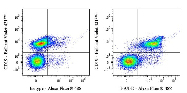
Flow cytometric analysis of C57BL/6 mouse splenocytes labeled with mouse I-A/I-E antibody at 1/500 dilution (1 μg) / (right panel) compared with a Rat IgG2b, κ Isotype Control / (left panel). Goat Anti-Rat IgG Alexa Fluor® 488 was used as the secondary antibody. Then cells were stained with CD19 - Brilliant Violet 421™ antibody separately. Total viable cells, as determined by Fixable Viability Dye 662 (S0B88806), were used for analysis. Flow cytometry and data analysis were performed using Agilent NovoCyte Quanteon and FlowJo™ software.
Invivo anti-Mouse I-A/I-E Recombinant mAb
Invivo anti-Mouse I-A/I-E Recombinant mAb
Price:
Regular price
$170 USD
Regular price
Sale price
$170 USD
Unit price
per
For shipping services or bulk orders, you may request a quotation.
Secure checkout with

View full details
Product Details
Product Details
Product Specification
| Host | Rat |
| Antigen | I-A/I-E |
| Synonyms | H-2 class II histocompatibility antigen, I-A beta chain; H2-Eb1 |
| Location | Membrane |
| Accession | P18468、 Q3U060 |
| Clone Number | M5/114.15.2 |
| Antibody Type | Rat mAb |
| Isotype | Rat IgG2b,k |
| Isotype Control | Invivo rat IgG2b isotype control, anti-keyhole limpet hemocyanin |
| Application | FCM, Functional assays, in vivo MHC II blockade |
| Reactivity | Ms |
| Positive Sample | C57BL/6 mouse splenocytes |
| Purification | Protein G |
| Concentration | 5 mg/ml |
| Purity | >95% (Determined by SDS-PAGE) |
| Endotoxin | <1EU/mg |
| Conjugation | Unconjugated |
| Physical Appearance | Liquid |
| Storage Buffer | PBS pH7.4, containing no preservative |
| Stability & Storage | 2 to 8 °C for 2 weeks under sterile conditions; |
Dilution
| application | dilution | species |
| FCM | 1:500 | Ms |
Background
I-A/I-E proteins are part of the Major Histocompatibility Complex (MHC) class II molecules, which are heterodimeric transmembrane glycoproteins expressed on antigen-presenting cells such as dendritic cells, macrophages, and B cells. These proteins are encoded by four genes located in the mouse MHC and are composed of α and β subunits. The I-A and I-E molecules are highly polymorphic and can form hybrid molecules in heterozygous individuals, contributing to the diversity of antigen presentation. They play a crucial role in presenting peptide antigens to CD4+ helper T cells, initiating adaptive immune responses. Additionally, MHC class II molecules can trigger intracellular signaling pathways in antigen-presenting cells, influencing their proliferation, maturation, and apoptosis.
Picture
Picture
FC