PD-L1/B7-H1 His Tag Protein, Mouse
PD-L1/B7-H1 His Tag Protein, Mouse
Price:
Regular price
$500 USD
Regular price
Sale price
$500 USD
Unit price
per
For shipping services or bulk orders, you may request a quotation.
Secure checkout with

View full details
Product Details
Product Details
Product Specification
| Species | Mouse |
| Synonyms | PD-L1/B7-H1 His Tag, Mouse |
| Accession | Q9EP73 |
| Amino Acid Sequence |
Phe19-Thr238, with C-terminal 8* His Tag FTITAPKDLYVVEYGSNVTMECRFPVERELDLLALVVYWEKEDEQVIQFVAGEEDLKPQHSNFRGRASLPKDQLLKGNAALQITDVKLQDAGVYCCIISYGGADYKRITLKVNAPYRKINQRISVDPATSEHELICQAEGYPEAEVIWTNSDHQPVSGKRSVTTSRTEGMLLNVTSSLRVNATANDVFYCTFWRSQPGQNHTAELIIPELPATHPPQNRTGGGSHHHHHHHH |
| Expression System | HEK293 |
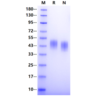
| Molecular Weight | 40-53kDa |
| Purity | >95% by SDS-PAGE |
| Endotoxin | <0.1EU/μg |
| Conjugation | Unconjugated |
| Tag | His Tag |
| Physical Appearance | Lyophilized Powder |
| Storage Buffer | PBS, pH7.4 |
| Reconstitution | Reconstitute at 0.1-1 mg/ml according to the size in ultrapure water after rapid centrifugation. |
| Stability & Storage | · 12 months from date of receipt, lyophilized powder stored at -20 to -80℃. · 3 months, -20 to -80℃ under sterile conditions after reconstitution. · 1 week, 2 to 8℃ under sterile conditions after reconstitution. · Please avoid repeated freeze-thaw cycles. |
| Reference |
1、Salih H R. et al. (2006) The role of leukemia-derived B7-H1 (PD-L1) in tumor-T-cell interactions in humans. Exp Hematol. 34(7): 888-894. 2、Wilcox R A. et al. (2009) B7-H1 (PD-L1, CD274) suppresses host immunity in T-cell lymphoproliferative disorders. Blood. 114(10): 2149-2158. 3、Ruggiero A. et al. (2009) Crystal structure of PD-L1, a ribosome inactivating protein from Phytolacca dioica L. leaves with the property to induce DNA cleavage. Biopolymers. 91(12): 1135-1142. |
Background
Programmed death ligand 1 ( PD-L1) belongs to the B7 series and is a 33-kDa type 1 transmembrane glycoprotein that contains 290 amino acids with Ig-V and IgC domains in its extracellular region. PD-L1 expression can be detected on hematopoietic cells including T cells, B cells, macrophages, dendritic cells (DCs), and mast cells, and non-hematopoietic healthy tissue cells including vascular endothelial cells, keratinocytes, pancreatic islet cells, astrocytes, placenta syncytiotrophoblast cells, and corneal epithelial and endothelial cells. PD-L1 is an essential immune checkpoint protein that binds to programmed death 1 (PD-1) on T-lymphocytes. Engagement of PD-1 by PD-L1 alters the activity of T cells in many ways, inhibiting T cell proliferation, survival, cytokine production, and other effector functions. T cell plays a critical role in killing cancer cells while the cancer cell exhibits immune escape by the expression of PD-L1. The binding of PD-L1 to PD-1 inhibits T cell proliferation and activity, leading to tumor immunosuppression.
Picture
Picture
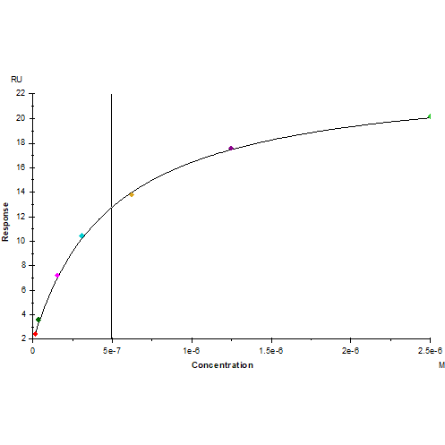
Bioactivity

PD-1 Fc Chimera,Mouse (Cat. No. UA010222) captured on Protein A Biosenor, can bind PD-L1/B7-H1 His Tag, Mouse (Cat. No. UA010223) with an affinity constant of 0.49μM as determined in SPR assay.
SDS-PAGE
1 μg (R: reducing conditions, N: non-reducing conditions).