Product Specification
| Host |
Rabbit |
| Antigen |
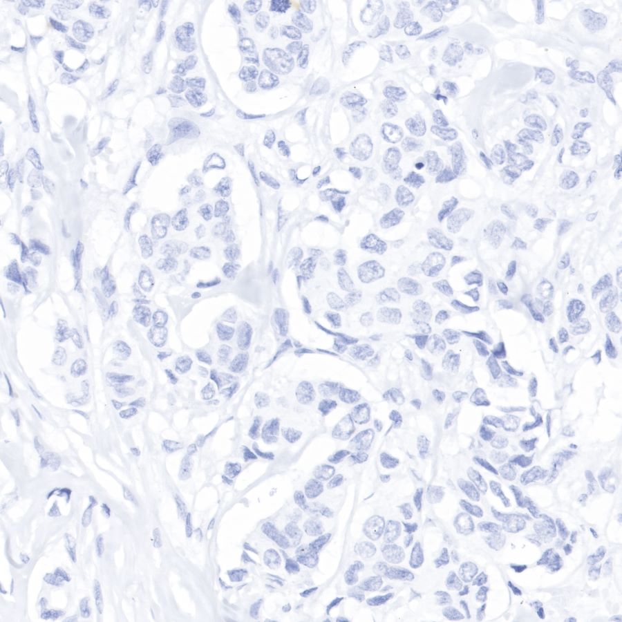
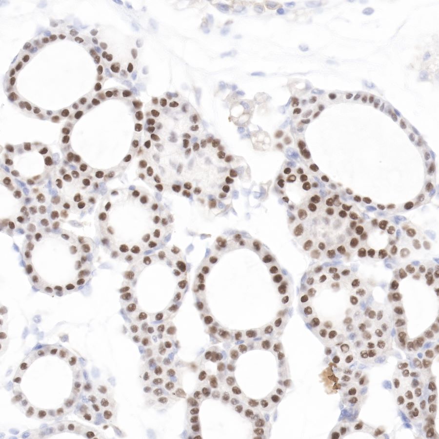
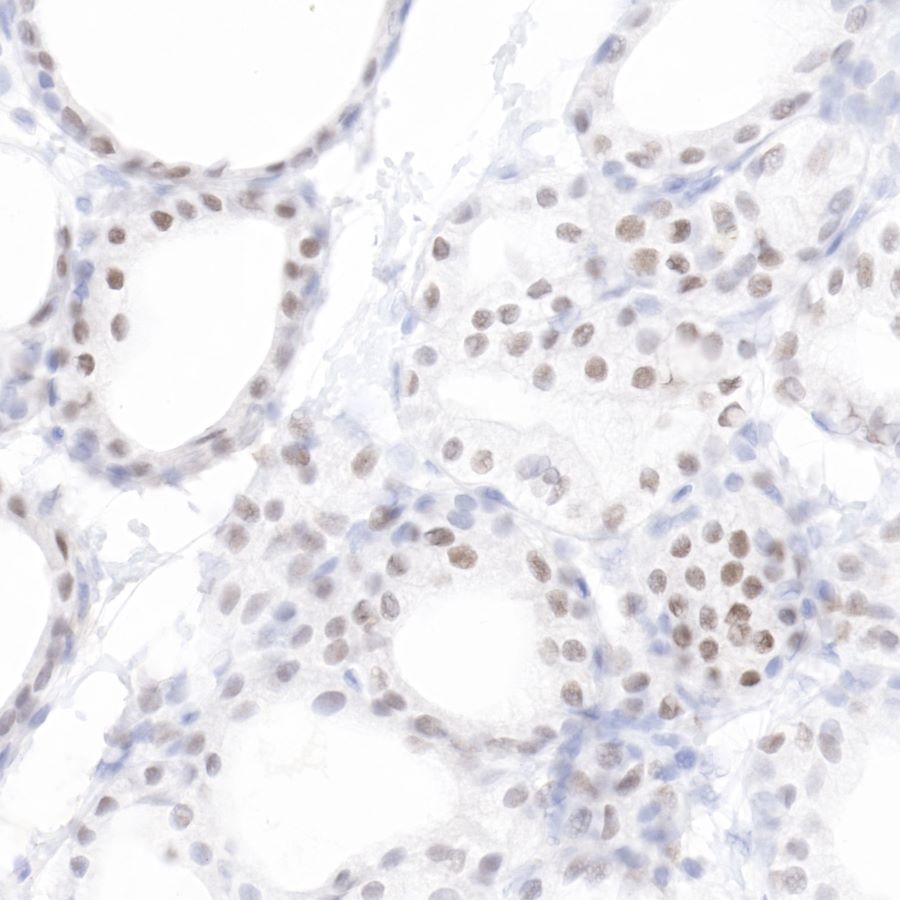
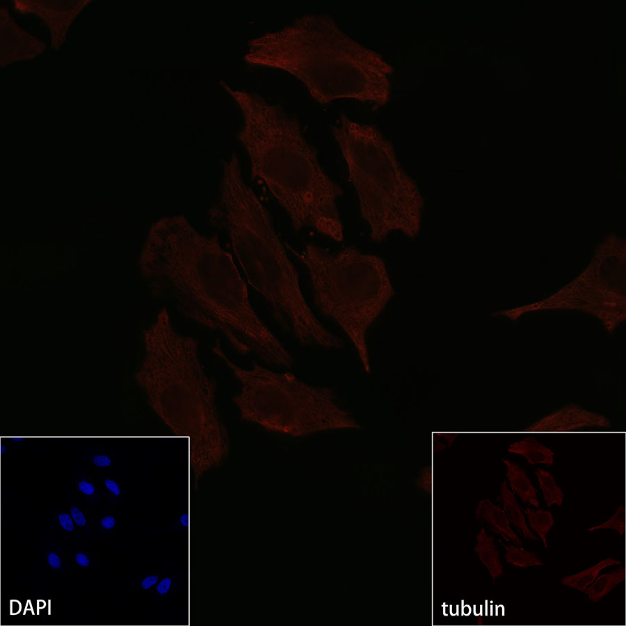
PAX8 |
| Immunogen |
Recombinant Protein |
| Location |
Nucleus |
| Accession |
Q06710 |
| Clone Number |
SDT-R027 |
| Antibody Type |
Rabbit mAb |
| Application |
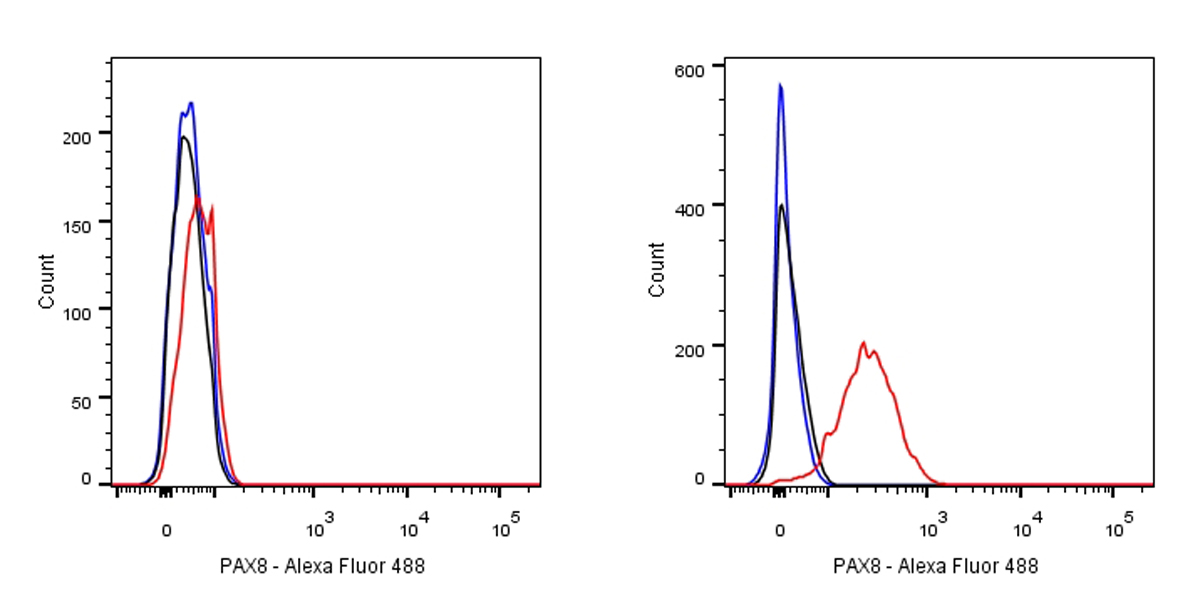
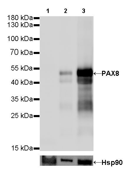
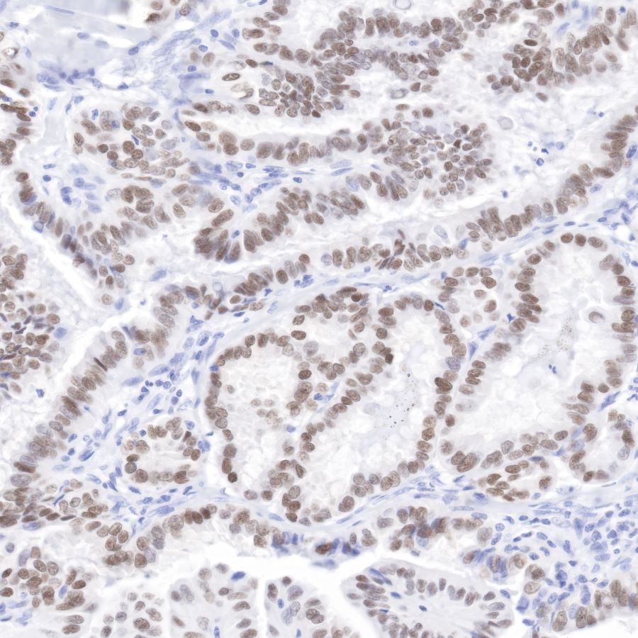
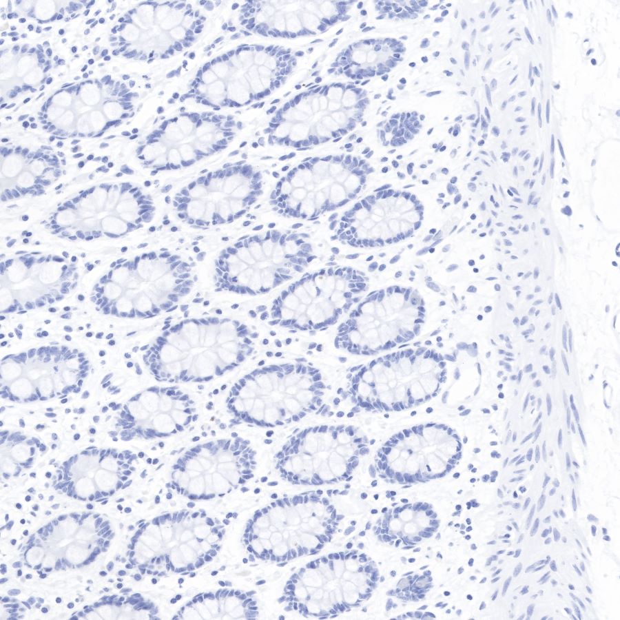
WB, IHC-P, ICC, FC |
| Reactivity |
Hu, Ms, Rt |
| Purification |
Protein A |
| Concentration |
2 mg/ml |
| Physical Appearance |
Liquid |
| Storage Buffer |
PBS, 40% Glycerol, 0.05%BSA, 0.03% Proclin 300 |
| Stability & Storage |
12 months from date of receipt / reconstitution, -20 °C as supplied |
Dilution
| application |
dilution |
species |
| IHC-P |
1:1000 |
null |
| FC |
1:250 |
null |
| ICC |
1:250 |
null |
| WB |
1:500 |
null |
Background
Paired box gene 8, also known as PAX8, is a protein which in humans is encoded by the PAX8 gene. This gene is a member of the paired box (PAX) family of transcription factors. Members of this gene family typically encode proteins which contain a paired box domain, an octapeptide, and a paired-type homeodomain. The PAX gene family has an important role in the formation of tissues and organs during embryonic development and maintaining the normal function of some cells after birth. PAX8 (and PAX2) is one of the important regulators of urogenital system morphogenesis.